Download
1 / 37
370 likes | 386 Views
The nervous and endocrine systems work together to coordinate body functions and maintain homeostasis. The endocrine system produces chemical messengers that circulate through the body, while the nervous system rapidly monitors and controls organ systems. This article discusses the functions of the nervous system, sensory input, integration and output, and the different components of the peripheral and central nervous systems.

E N D
Mechanisms forco-ordinating homeostasisin mammals Nervous & Endocrine Systems
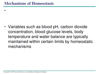
Nervous and Endocrine Systems • The nervous and endocrine systems work together to coordinate the actions of all other systems of the body to produce behavior and maintain homeostasis. • Theendocrine system produces chemical messengers that are transported through the circulatory system. It requires seconds, minutes or hours. • The nervous system is more rapid, requiring only thousandths of a second. • In general, the endocrine system is in charge of body processes that happen slowly, such as cell growth. • Faster processes like breathing and body movement are monitored by the nervous system.
Functions of nervous system • Receive sensory input from internal and external environments. • Integrate the input, and respond to stimuli.
Nervous System Sensory Input • Receptors are parts of the nervous system that sense changes in the internal or external environments. • Sensory input can be in many forms, including pressure, taste, sound, light, blood pH, or hormone levels, that are converted to a signal and sent to the brain or spinal cord. Integration and Output • In the sensory centers of the brain or in the spinal cord, the barrage of input is integrated and a response is generated. • The response, a motor output, is a signal transmitted to organs than can convert the signal into some form of action, such as movement, changes in heart rate, release of hormones, etc.
Nervous System • The nervous system monitors and controls almost every organ system through a series of positive and negative feedback loops. • It is divided into two parts: • Central Nervous System: brain and spinal cord • Peripheral Nervous System: all other nerve tissue. Can be further divided based on the function of its parts.
NERVOUS SYSTEM PERIPHERAL CENTRAL Brain Spinal Cord Somatic Autonomic Sensory Motor Sympathetic Parasympathetic
Peripheral Nervous System • Contains only nerves and connects the brain and spinal cord (CNS) to the rest of the body. • Axons and dendrites are surrounded by a white myelin sheath. • Cell bodies are in the central nervous system (CNS) or ganglia (collections of nerve cell bodies). • Two main components of the PNS: • sensory (afferent) pathways that provide input from the body into the CNS. • motor (efferent) pathways that carry signals to muscles and glands (effectors). • Most sensory input carried in the PNS remains below the level of conscious awareness. Input that does reach the conscious level contributes to perception of our external environment. • There are two major subdivisions of the PNS motor pathways: the somaticand the autonomic.
Peripheral Nervous System Somatic Nervous System (voluntary responses) • Includes all nerves controlling the muscular system and external sensory receptors. • External sense organs (including skin) are receptors. • Muscle fibers and gland cells are effectors. • Sensory input from the PNS is processed by the CNS and responses are sent by the PNS from the CNS to the organs of the body. • Motor neurons of the somatic system are distinct from those of the autonomic system. Inhibitory signals cannot be sent through the motor neurons of the somatic system.
Peripheral Nervous System Autonomic Nervous System (Involuntary Responses) • Consists of motor neurons that control internal organs i.e. muscles in the heart, the smooth muscle in internal organs such as the intestine, bladder, and uterus. • Has two subsystems: • Sympathetic Nervous System is involved in the fight or flight response. • Parasympathetic Nervous System is involved in relaxation. • Each of these subsystems operates in the reverse of the other (antagonism). • Both systems innervate the same organs and act in opposition to maintain homeostasis. • For example: when you are scared the sympathetic system causes your heart to beat faster; the parasympathetic system reverses this effect.
Central Nervous System • Is composed of the brain and spinal cord. • Surrounded by bone-skull and vertebrae. Fluid and tissue also insulate the brain and spinal cord. • The central nervous system receives impulses from sensory nerves, coordinates responses and directs the action of effectors. • Brain • Coordinates actions and responses, both voluntary and involuntary • Spinal Cord • Carries messages from the brain to motor or autonomic nerves, and to the brain from sensory nerves. • Directs the response in a reflex arc.
What is a reflex arc? • The brain generally coordinates responses to information from receptors. • Sometimes however the body cannot wait for the transmission of information from the spinal cord to the brain and back from the brain to an effector. This is where the reflex arc comes in…. • The reflex arc is an automatic, involuntary reaction to a stimulus. • Examples of reflex arcs include balance, the blinking reflex, and the stretch reflex.
Example of a reflex arc Touching a sharp object • Receptor detects pain • Sensory neurons carry signal to spinal cord • Interneurons in the spinal cord directly stimulate effectors (e.g. skeletal muscle) using motor neurons • Spinal interneurons also transmit pain signal to the brain This is why you drop the object and then a moment later feel the pain • Brain then coordinates the rest of your voluntary and involuntary responses to pain.
Brain • Cerebral cortex • Receives sensory information, links sensory and motor functions • Cerebellum • Coordinates posture and balance • Thalamus • Relays sensory information from the brainstem spinal cord and cerebellum to the cerebral cortex • Hypothalamus • Control centre for most homeostatic mechanisms, involuntary nervous responses and hormonal responses • Brain stem • Links brain and spinal cord, controls some automatic functions and reflexes
Nervous Tissue • Nervous tissue is composed of two main cell types: neurons and glial cells. • Neurons transmit nerve messages. • Glial cells are in direct contact with neurons and often surround them.
Neurons • The neuron is the functional unit of the nervous system. • Humans have about 100 billion neurons in their brain alone! • Although they vary in size and shape, all neurons have three parts: • Dendrites receive information from another cell and transmit the message to the cell body. • Cell body contains the nucleus, mitochondria and other organelles typical of eukaryotic cells. • Axon conducts messages away from the cell body. Axon terminals form synapses with other neurons or with a muscle or a gland.
Three types of neurons • Sensory neurons typically have a long dendrite and short axon, and carry messages from sensory receptors to the central nervous system. Also known as affector neurons. • Motor neurons have a long axon and short dendrites and transmit messages from the central nervous system to the muscles (or to glands). Also known as effector neurons. • Interneuronsare found only in the central nervous system where they connect neuron to neuron. Also known as connecting neurons.
The Nerve Message - Electrical • In a resting nerve (one that is not responding to stimulus), a small difference exists between the electrical charge on the inside and outside its cell membrane. The outside of the cell membrane of the axon is positive compared with the inside. • Stimuli of various kinds can activate neurons so that they transmit nerve impulses along their axons. Such a nerve cell is said to be ‘excited’. • Nerve impulses involve changes in the charge across the axon membranes. • As the impulse moves along the axon, a change occurs in the permeability of the membrane so that positive ions move into the cell. • This results in the outside of the membrane becoming negative compared with the inside. The change in permeability travels along the neuron. • After a nerve impulse has been transmitted by a neuron, the original distribution of ions across the cell membrane is restored.
Myelin sheath • Acts as insulator to minimize metabolic expense while maintaining rapid conduction of electrical impulse through neurons. • Sheaths are formed by glial cells: • oligodendrocytes in the central nervous system • Schwann cells in the peripheral nervous system. • The myelin sheath in peripheral nerves normally runs along the axon in sections about 1 mm long, punctuated by unsheathed nodes of Ranvier which contain a high density of voltage-gated ion channels. • An action potential is conducted more rapidly along a myelinated axon because it ‘jumps’ from one node to the next.
The Nerve Message - Chemical • Neurons communicate with one another via synapses, where the axon terminal of one cell impinges upon a dendrite or soma of another (or less commonly to an axon). • The human brain has a huge number of synapses. Estimates vary for an adult, ranging from 100 to 500 trillion synapses. • The space between two cells is known as the synaptic cleft. For signals to cross the synaptic cleft neurotransmitters are required. • The time for neurotransmitter action is between 0.5 and 1 millisecond. • Acetylcholine is an example of a neurotransmitter, as is norepinephrine, although each acts in different responses.
How do neurotransmitters work? • Neurotransmitters are stored in small synaptic vesicles clustered at the tip of the axon. • Arrival of the action potential causes some of the vesicles to move to the end of the axon and discharge their contents into the synaptic cleft. • Released neurotransmitters diffuse across the cleft, and bind to receptors on the other cell's membrane, causing ion channels on that cell to open and prompting transmission of the message along that cell’s membrane. • Some neurotransmitters cause an action potential, others are inhibitory. • Once in the cleft, neurotransmitters are active for only a short time. • Enzymes in the cleft inactivate the neurotransmitters. Inactivated neurotransmitters are taken back into the axon and recycled.
Toxins • Many animal toxins act on the nervous system, particularly at neuro-muscular synapses. • Some prevent the passage of nerve impulses along a nerve. • Others act on one or both sides of the neuro-muscular synapse.
Endocrine System • The foundations of the endocrine system are the hormones and glands. • As the body's chemical messengers,hormones transfer information and instructions from one set of cells to another. • Although many different hormones circulate throughout the bloodstream, each one affects only the cells that are genetically programmed to receive and respond to its message. • A gland is a group of cells that produces and secretes, or gives off, chemicals. It selects and removes materials from the blood, processes them, and secretes the finished chemical product for use somewhere in the body. • Some types of glands release their secretions in specific areas. For instance, exocrineglands, such as the sweat and salivary glands, release secretions in the skin or inside of the mouth. • Endocrine glands, on the other hand, release more than 20 major hormones directly into the bloodstream where they can be transported to cells in other parts of the body.
Endocrine System • The major glands that make up the human endocrine system are: • Hypothalamus • Pituitary • Thyroid • Parathyroids • Adrenals • Pineal body • Reproductive glands (ovaries and testes) • The pancreas is also part of this hormone-secreting system, even though it is also associated with the digestive system because it also produces and secretes digestive enzymes. • Some non-endocrine organs - such as the brain, heart, lungs, kidneys, liver, thymus, skin, and placenta - also produce and release hormones.
Hypothalamus • The hypothalamusis the primary link between the endocrine and the nervous system • It is a collection of specialized cells that is located in the lower central part of the brain. • Nerve cells in the hypothalamus control the pituitary gland by producing chemicals that either stimulate or suppress hormone secretions from the pituitary.
Pituitary Glands • Located at the base of the brain just beneath the hypothalamus. • Considered the most important part of the endocrine system because it makes hormones that control several other endocrine glands. • The production and secretion of pituitary hormones can be influenced by factors such as emotions and seasonal changes. • To accomplish this, the hypothalamus relays information sensed by the brain (such as environmental temperature, light exposure patterns, and feelings) to the pituitary. • Secretes endorphins,chemicals that act on the nervous system to reduce sensitivity to pain. • Secretes hormones that signal the ovaries and testes to make sex hormones. • Controls ovulation and the menstrual cycle in women.
Pituitary Glands • The pituitary is divided into two parts: the anterior lobe and the posterior lobe. • The anterior lobe regulates the activity of the thyroid, adrenals, and reproductive glands. It produces: • growth hormone: stimulates the growth of bone and other body tissues and plays a role in the body's handling of nutrients and minerals • prolactin: activates milk production in women who are breastfeeding • thyrotropin:stimulates the thyroid gland to produce thyroid hormones • corticotropin:stimulates the adrenal gland to produce certain hormones • The posterior lobe of the pituitary releases antidiuretichormone, which helps control body water balance through its effect on the kidneys and urine output; and oxytocin which triggers the contractions of the uterus that occur during labor.
Thyroid and Parathyroid Glands Thyroid • Located in the front part of the lower neck, and shaped like a bowtie or butterfly. • Produces the thyroid hormones thyroxine and triiodothyronine. • These hormones control the rate at which cells burn fuels from food to produce energy. As the level of thyroid hormones increases in the bloodstream, so does the speed at which chemical reactions occur in the body. • Thyroid hormones also play a key role in bone growth and the development of the brain and nervous system in children. • The production and release of thyroid hormones is controlled by thyrotropin,which is secreted by the pituitary gland. Parathryoids • Attached to the thyroid are four tiny glands that function together called the parathyroids. • They release parathyroid hormone, which regulates the level of calcium in the blood with the help of calcitonin, which is produced in the thyroid.
Adrenal and Pineal Glands Adrenal Glands • Adrenal glands are triangular in shape. • There are two adrenal glands, one on top of each kidney. • The adrenal glands have two parts, each of which produces a set of hormones and has a different function. • The outer part, the adrenal cortex, produces hormones called corticosteroids that influence or regulate salt and water balance in the body, the body's response to stress, metabolism, the immune system, and sexual development and function. • The inner part, the adrenal medulla, producescatecholamines such as epinephrine. Also called adrenaline, epinephrine increases blood pressure and heart rate when the body experiences stress. Pineal Gland • Also called the pineal body. • Located in the middle of the brain. • It secretes melatonin, a hormone that may help regulate the wake-sleep cycle.
Gonads • The gonads are the main source of sex hormones. • Male Gonads – The Testes • Located in the scrotum • Secrete hormones called androgens, the most important of which is testosterone. • These hormones regulate body changes associated with sexual development, including enlargement of the penis, the growth spurt that occurs during puberty, and the appearance of other male secondary sex characteristics such as deepening of the voice, growth of facial and pubic hair, and the increase in muscle growth and strength. • Working with hormones from the pituitary gland, testosterone also supports the production of sperm by the testes. • Female Gonads – The Ovaries • Located in the pelvis • They produce eggs and secrete the female hormones estrogen and progesterone. • Estrogen is involved in the development of female sexual features such as breast growth, the accumulation of body fat around the hips and thighs, and the growth spurt that occurs during puberty. • Both estrogen and progesterone are also involved in pregnancy and the regulation of the menstrual cycle.
Pancreas • The pancreasproduces (in addition to others) two important hormones, insulin and glucagon. • They work together to maintain a steady level of glucose, or sugar, in the blood and to keep the body supplied with fuel to produce and maintain stores of energy.
Endocrine System and Negative Feedback • When hormone levels reach a certain normal or necessary amount, further secretion is controlled by important body mechanisms to maintain that level of hormone in the blood. • Regulation of hormone secretion may involve the hormone itself or another substance in the blood related to the hormone. • For example, if the thyroid gland has secreted adequate amounts of thyroid hormones into the blood, the pituitary gland senses the normal levels of thyroid hormone in the bloodstream and adjusts its release of thyrotropin, the pituitary hormone that stimulates the thyroid gland to produce thyroid hormones. • Another example is parathyroid hormone, which increases the level of calcium in the blood. When the blood calcium level rises, the parathyroid glands sense the change and decrease their secretion of parathyroid hormone. This turnoff process is called a negative feedback system.
Elimination of hormones • Hormones do not last indefinitely. • Once they have delivered their signal to target cells and the desired effect has occurred, they are no longer required. • They are degraded by cell enzymes and excreted via the kidneys or faeces.