Download
1 / 39
390 likes | 645 Views
Minor Brain Trauma: Pathology, Imaging, and Clinical Aspects. Donald W Chakeres MD, FACR Professor of Radiology Department of Radiology The Ohio State University Columbus, Ohio, USA. XIX Symposium Neuroradiologicum, Bologna, Italy 2010. Thanks to the Collaborators. Seongjin Choi, PhD

E N D
Minor Brain Trauma: Pathology, Imaging, and Clinical Aspects Donald W Chakeres MD, FACR Professor of Radiology Department of Radiology The Ohio State University Columbus, Ohio, USA XIX Symposium Neuroradiologicum, Bologna, Italy 2010
Thanks to the Collaborators Seongjin Choi, PhD Postdoctoral Fellow Department of Radiology John D. Corrigan, PhD Professor Department of Phys. Med. and Rehab. Department of Psychology Jennifer A. Bogner, PhD Associate Professor Department of Phys. Med. and Rehab. W Jerry Mysiw, MD Professor and Vice Chair Department of Phys. Med. and Rehab. Department of Psychology S. Sammet MD, PhD Assistant Professor Department of Radiology Dustin Cunningham Student Research Assistant Department of Radiology Devin F. Prior Student Research Assistant Department of Radiology Cherian R Zachariah, BS Graduate Research Associate Department of Radiology Michael V. Knopp, MD, PhD Professor and Vice Chair Department of Radiology Petra Schmalbrock, PhD Associate Professor Department of Radiology
Goals • Preview of the presentation • Define minor head trauma • Clinical findings/ spectrum • Pathology, animal and human • Imaging findings, focusing on 7 T MRI • Conclusions
Disclaimer for the Presentation • If you are looking for great imaging findings that will impact your routine practice, you are going to be disappointed • Minor head trauma by definition usually does not have routine imaging findings • Still a very important topic since it is a common indication for imaging • Advanced imaging does have the potential to become the biomarker measure
Significance - Incidence • Head trauma is extremely common • Minor head trauma is commonly followed by repeated or major head trauma • In America there are over 5 million with long term disability as a result of head trauma • The incidence of minor head trauma is essentially everyone • It is a very serious problem dwarfing many other medical issues
Treatment • There is no known effective therapy • Most therapy is just to wait and see if the symptoms improve, frequently they do • The treatment for stress and trauma are frequently identical • If you cannot measure the injury directly you cannot measure if your therapy is effective either
Definitions • The classification is not well defined for imaging or clinical measures • Glasgow Coma Scale, not good for minor head injuries, better for serious injuries
Definition: Minor Head Trauma American Academy of Neurology • Grade 1: transient confusion with no loss of consciousness, and symptoms resolve in less than 15 minutes • Grade 2: symptoms last for more than 15 minutes • Grade 3: loss of consciousness
Definition Minor Head Trauma: Imaging • Normal routine CT and MRI
Clinical Symptoms • Loss of consciousness • Head ache • Confusion • Dizzy • Inability to concentrate • Does not recall the event • Change in personality • Executive function impairment
Clinical Symptoms: Compounded because the findings overlap with many other issues • Post traumatic stress syndrome • Depression • Drug dependence • Workman’s compensation incentives
Major Trauma Pathology: • Major brain injuries much better understood because of excellent pathologic material • Disruption without physical shearing of the neurofilaments • Animal studies show “ball” like swelling of axons suggesting separation, but without gross visible focal parenchymal injury, analogous to MS and normal appearing white matter • Stretching and tearing of neurons occur at the instant of trauma Minor Trauma Pathology: Povlishock JT, Becker DP, et al, Axonal change in minor head Injury, Journal of Neuropathology and Experimental Neurology
Minor Trauma Pathology • Wide spread damage to the white matter need not be associated with hemorrhage, contusion, or laceration • This is why advanced imaging may be very important Nevin, Norman, Neuropahtological changes in white matter following head trauma, Journal of Neuropathology and Experimental Neurology 1967
Palmgren silver impregnation of swollen Purkinje cell dendrites (arrows) in cerebellar molecular layer Kors E, et al, Delayed cerebral edema and fatal coma after minor head trauma: role of the CACNA1A calcium channel subunit gene and relationship with familial hemiplegic migraine, Ann Neurol 2001:49:753-760
Minor Trauma Pathology • Loss of consciousness may the clinical marker of the “tearing” of the neurons • The brainstem foci may well account for loss of consciousness • Location can be “anywhere”, but classic anatomic distribution • Animal studies show injuries to the inferior colliculus, pons and dorsolateral medulla • Axonal injury is highly under estimated Jane J, et al, Axonal degeneration induced by experimental noninvasive minor head injury Gentleman, SM, Roberts GW, et al , Axonal injury: a universal consequence of Fatal closed head injury, Acta Neuropathol 1995:89:537-543
Major Trauma Pathology: Midbrain Lesion and Prognosis • Midbrain lesions found in 23 of 35 autopsied, head injury subjects • Abnormal evoked potential before death • Midbrain damage almost always associated with major hemispheric injury Rosenblum, W, et al, Midbrain lesion: frequent and significant prognostic feature in closed head injury Neurosurgery, Congress of Neurologic Surgeons
Genetic Predispositions • ApoE is a genetic marker that is associated with Alzheimer’s disease • An error in “healing” the brain after injury • The risk of significant post traumatic disability is increased for subjects with the same ApoE genes as patients at risk for Alzheimer’s disease • Beta-amyloid deposits are found with the apolipoprotineE (apoE)-ε4 allele. β‐Amyloid (Aβ)42(43), Aβ42, Aβ40 and apoE immunostaining of plaques in fatal head injury K. Horsburgh, G. M. Cole, F. Yang, M. J. Savage B. D. Greenberg, S. M. Gentleman, D. I. Grahamand J. A. R. Nicoll Association of apolipoprotein E polymophism with outcome after head injury, The Lancet volume 350, issue 9084, 1069-1071
Blast Injuries, Minor or Major • 90% improve with no therapy • Can occur without any head motion, classic acceleration- deceleration • The blast waves can go through the thinner bone skull regions such as the orbit and still lead to distortion/ injury of the brain • Usually more than one blast needed before any clinical symptoms
Imaging of Major Head Trauma • Subdural hematoma • Epidural hematoma • Subarachnoid hemorrhage • Brain contusions • Diffuse axonal injuries • Brain lacerations • Depressed skull fracture • Vessel disruption, spasm, dissection, aneurym
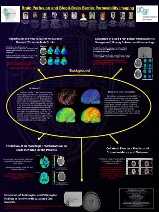
Susceptibility Weighted Imaging • Technique developed by Haacke/ et al • More commonly available now, and standard on some systems • Uses a image processed combination of phase and magnitude data • Very sensitive to deoxyhemoglobin since it has strong paramagnetic properties that accelerate the local phase • Good for the veins and hemorrhage
Susceptibility Weighted Imaging • Much better than simple gradient echo imaging for subtle blood products • Patients with “normal CT and MRI” may have many lesions by SWI • Seen as low “signal” regions in typical locations for shear injuries and trauma
MRI Spin Echo: Brain Contusions FLAIR T2
SWI: Brain Contusions SWI T2
Diffusion Imaging of Trauma • Diffusion imaging should be a good technique for a subtle injury since it is sensitive to the integrity of the axons • FA and ADC values may be abnormal with brain injury • Diffusion tensor/ tract imaging, DTT, should even be more sensitive
7 T Research Study of Trauma • Data comparable to 3 T data, but higher resolution • Fractional anisotropy, FA, values lower in corpus callosum, CC, compared to controls • Healthy normal subjects showed changes of FA with age, FA decreases with increasing age • Tractography of CC showed fewer and less well defined tracts in the trauma group
7T Tractography of Normal and Post Traumatic Subjects Normal control Post Traumatic Subject
7T DTI in Chronic Mild Traumatic Brain Injury: Assessment of the Superior Longitudinal Fasciculus and Cingulum Bundle Dustin T. Cunningham, Seongjin Choi, John D. Corrigan, Jennifer Bogner, W. Jerry Mysiw,Cherian R. Zachariah, Michael V. Knopp and Petra Schmalbrock The Ohio State University Department of Radiology Wright Center of Innovation in Biomedical Imaging
Methods • Subjects • 7 chronic mild TBI with normal conventional MRI (ages 22-60) • (Glasgow Coma Scale GCS 13-15) • 10 approx. age matched healthy (22-56) • Imaging • 7T (Philips, Achieva) • 16-channel receive (Nova Medical) • SS-SE-EPI DTI, TR/TE = 5126/75 ms • voxel: 1.6×1.6×3.2 mm3 • b = 0, 1000 s/mm2 • 6 b-directions, 3 high b averages • SENSE-factor = 5 • DTI scan time: 2min, 24sec • 3D SWI, voxel: .40×.68×1.6 mm3 • flip angle = 5, scan time: 5min, 4sec
Cingulum Bundles 7T Identification Philips FiberTrak Wakana et al. NeuroImage 2007
CB Tractography Scoring 3 2 1 DTI Studio – H. Jiang and S. Mori, Johns Hopkins University
CB Tractography Scoring Control TBI Observer 1 Observer 2
Summary • Visual tractography differences can be detected in chronic mild TBI compared with controls with 7T • Quantitative FA values were lower in chronic mild TBI than controls • Despite B0 and B1 inhomogeneity, 7T DTI consistent with 3T and 1.5T studies of mild TBI
References • Inglese M, Makani S, Johnson G, Cohen BA, Silver JA, Gonen O, and Grossman RI, Diffuse Axonal Injury in Mild Traumatic Brain Injury: A Diffusion Tensor Imaging Study, Journal of Neurosurgery 103:298-303, 2005 • Kraus MF, Susmaras T, Caughlin BP, Walker CJ, Sweeney JA, and Little DM, White Matter Integrity and Cognition in Chronic Traumatic Brain Injury: A Diffusion Tensor Imaging Study, Brain 130:2508-2519, 2007 • Kumar R, Husain M, Gupta RK, Hasan KM, Haris M, Agarwal AK, Pandey CM, and Narayana PA, Serial Changes in the White Matter Diffusion Tensor Imaging Metrics in Moderate Traumatic Brain Injury and Correlation with Neuro-Cognitive Function, Journal of Neurotrauma 26:481-495, April 2009 • Li TQ, van Gelderen P, Merkle H, Talagala L, Koretsky AP, Duyn J, Extensive heterogeneity in white matter intensity in high-resolution T2*-weighted MRI of the human brain at 7T, NeuroImage 32, 1032-1040, 2006 • Sugiyama K, Kondo T, Higano S, Endo M, Watanabe H, Shindo K, Izumi SI, Diffusion tensor imaging fiber tractography for evaluating diffuse axonal injury, Brain Injury, 21: 413-419, 2007
Conclusion: • “Minor” head trauma is a major medical issue • Pathology has shown many important findings not commonly identified otherwise • Some patients are at a genetic risk • MR imaging may be the future of an accurate biomarker for brain injury • “Normal” imaging may not be normal • Advanced techniques are much more sensitive for imaging findings of TBI
Thanks to the organizers of the meeting and have a great day in lovely Italy