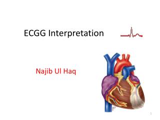
ECGG Interpretation
370 likes | 549 Views
ECGG Interpretation. Najib Ul Haq. ECG. ECG Basics Normal Sinus Rhythm How to Analyze a Rhythm Heart Arrhythmias Diagnosing a Myocardial Infarction Advanced 12-Lead Interpretation. Normal Cardiac Current Flow. Basics of Current movement.

ECGG Interpretation
E N D
Presentation Transcript
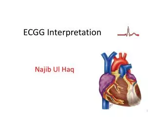
ECGG Interpretation Najib Ul Haq
ECG • ECG Basics • Normal Sinus Rhythm • How to Analyze a Rhythm • Heart Arrhythmias • Diagnosing a Myocardial Infarction • Advanced 12-Lead Interpretation
Basics of Current movement • Current + Electrode • Electrode Current
Normal Impulse Conduction Sinoatrial node AV node Bundle of His Bundle Branches Purkinje fibers
Impulse Conduction & the ECG Sinoatrial node AV node Bundle of His Bundle Branches Purkinje fibers
The PR Interval Atrial depolarization + Delay in AV junction (AV node/Bundle of His) (Delay allows time for the atria to contract before the ventricles contract)
The “PQRST” • P wave - Atrial depolarization • QRS- Ventricular depolarization • T wave- Ventricular repolarization
Einthoven’s Triangle Lead I - + extends from the right to the left arm - Lead III extends from the left arm to the left foot Lead II extends from the right arm to the left foot +
12-Lead ECG Rhythm Strip Diagnosing MI To diagnose a myocardial infarction you need to go beyond looking at a rhythm strip and obtain a 12-Lead ECG.
The 12-Lead ECG • The 12-Lead ECG sees the heart from 12 different views. • Therefore, the 12-Lead ECG helps you see what is happening in different portions of the heart. • The rhythm strip is only 1 of these 12 views.
The 12-Leads The 12-leads include: • 3 Limb leads (I, II, III) • 3 Augmented leads (aVR, aVL, aVF) • 6 Precordial leads (V1- V6)
Views of the Heart Lateral portion of the heart Some leads get a good view of the: Anterior portion of the heart Inferior portion of the heart
ST Elevation One way to diagnose an acute MI is to look for elevation of the ST segment.
A Normal 12 Lead ECG 2004 Anna Story
ST Elevation (cont) Elevation of the ST segment (greater than 1 small box) in 2 leads is consistent with a myocardial infarction.
Left Main Coronary Artery • Branches quickly into the LAD & LCX. Involves almost 2/3 of the heart muscle primarily anterior • Right Coronary Artery (RCA)The RCA supplies blood to the bottom (inferior) portionand part of the back (posterior) portion of the left ventricle. The posterior portion of the septum is also supplied with blood from the RCA. • SA Node 55% • AV Node 90% • AV Blocks • Left Anterior Descending Branch (LAD)The LAD supplies blood to the front (anterior) portion of the left ventricle, apical including most of the anterior portion of the septum separating the ventricles. • Bundle Branch Block, AMI, CHF • Left Circumflux Branch (LCX)The LCX supplies blood to the left side (lateral) portion and the back (posterior) portion of the left ventricle. • SA Node 45% • AV Node 10% • Lateral & posterior MI
Lateral portion of the heart Anterior portion of the heart Inferior portion of the heart MI Locations Look again at this picture of the heart.
Lateral portion of the heart Anterior portion of the heart Inferior portion of the heart MI Locations Look again at this picture of the heart.
Anterior View of the Heart The anterior portion of the heart is best viewed using leads V1- V4.
Anterior MI Remember the anterior portion of the heart is best viewed using leads V1- V4. Limb Leads Augmented Leads Precordial Leads
Anterior Wall MI View of Anterior Heart Wall • Leads V3, V4 • Looks at anterior heart wall • Looks from the left anterior chest
Lateral MI So what leads do you think the lateral portion of the heart is best viewed? Leads I, aVL, and V5- V6 Limb Leads Augmented Leads Precordial Leads
Lateral Wall MI View of Lateral Heart Wall • Leads I and aVL • Looks at lateral heart wall • Looks from the left arm toward heart *Sometimes known as High Lateral*
Inferior MI Now how about the inferior portion of the heart? Leads II, III and aVF Limb Leads Augmented Leads Precordial Leads
Inferior Wall MI View of Inferior Heart Wall • Leads II, III, aVF • Looks at inferior heart wall • Looks from the left leg up
Putting it all Together Now, where do you think this person is having a myocardial infarction?
Inferior Wall MI This is an inferior MI. Note the ST elevation in leads II, III and aVF.
Inferior Wall MI This is an inferior MI. Note the ST elevation in leads II, III and aVF.
Putting it all Together How about now?
Anterolateral MI This person’s MI involves both the anterior wall (V2-V4) and the lateral wall (V5-V6, I, and aVL)!