Download
1 / 58
580 likes | 674 Views
Explore the functions, layers, and accessory structures of the skin. Learn about sebaceous and sweat glands, hair, nails, and dermatology. Discover common skin pathologies like acne vulgaris, comedo, and seborrhea.

E N D
Skin: The Integumentary System
Functions • Provides a barrier against hazardous materials and pathogens • Major receptor for the sense of touch • Waterproofs the body and prevents fluid loss • Helps the body synthesize vitamin D from ultraviolet light • Integument means “covering”
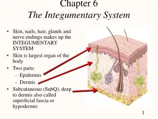
Structures • Skin • Largest organ of the body • Approx 5 kg in the average human • Dermat/o, cutane/o mean skin • Made up of two distinct tissue layers and an underlying layer of fatty tissue
Structures • Epidermis • Outermost layer of skin • Consists of stratified squamous epithelium • Top layers are flat overlapping cells that contain soft keratin(fibrous, water-repellant protein) – form the waterproof barrier
Structures • Lower layer is the basal layer – produces the epithelial cells that get pushed toward the surface where they die and become filled with keratin • The dead cells slough off so new cells can take their place • The basal layer also contains melanocytes that produce melanin (the dark brown pigment that determines skin colour, produces freckles, and protects against ultraviolet light)
Structures • Dermis • AKA corium • Contains connective tissue, blood and lymph vessels, nerve fibers • Collagen– tough, flexible, fibrous protein • Mast Cells – respond to injury, infection, or allergy by releasing substances like histamine and heparin • Provides a living substrate for the epidermis, and contains the accessory structures, such as hair follicles, glands, and nerve endings
Structures • Subcutaneous Layer • Connects the skin loosely to the underlying muscles • Consists of loose connective tissue and adipose tissue • Subcutaneous means “below the skin” • Provides a cushion against shock, and insulation against temperature changes
Accessory Structures • Sebaceous Glands • Associated with hair follicles • Except on palms of hands and soles of feet (most concentrated in the scalp) • Secrete sebum – an oily substance which lubricates the skin and discourages bacterial growth (it is slightly acidic) • Also helps to prevent water loss
Accessory Structures • Sweat Glands • AKA sudoriferous glands • Tiny, coiled glands that secrete perspiration (sweat) to the body surface • Most numerous on the palms of the hands, soles of the feet, forehead, armpits • Perspiration is composed primarily of water and some salt and waste products • Functions to excrete excess water and cool the body • Hidr/o means sweat • Hidrosis is the production of sweat
Accessory Structures • Eccrine Sweat glands • Open onto the skin surface and secrete through openings called pores • Most numerous type
Accessory Structures • Apocrine Sweat glands • Open into hair follicles • Most numerous in armpit and external genitalia • Substance secreted also contains proteins and fatty acids which give it a yellow colour • Respond to pain, emotional stress, sexual arousal, etc
Accessory Structures • Hair (trich/o, pil/o) • Columns made of dead protein cells filled with hard keratin • Melanocytes in the follicle produce melanin which colours the hair • Follicle: sac that holds the root of the hair (its shape determines whether hair will be straight or curly) • Arrector pili – tiny muscle fibers that contract to make the hair stand erect (reduces heat loss through the skin)
Accessory Structures • Nails • Thin plates of keratinized cells that protect the end of the fingers and toes • Nail Body: translucent, closely molded to underlying tissues
Accessory Structures • Nail Bed: joins the nail body to the underlying tissue and nourishes the nail • Very vascularized – gives the nail its pink colour • Root: fastens the nail to the finger or toe by fitting it into a groove in the skin
Accessory Structures • Free Edge: portion of the nail body not attached to the nail bed (white portion of the nail) • Lunula: pale, half-moon shaped region at the root of the nail • Part of the nail body that covers the matrix (active area where new cells form)
Accessory Structures • Cuticle: Narrow band of epidermis attached to the surface of the nail just in front of the root • Protects the new cells as they grow • Onych/o, ungu/o mean nail
Medical Specialties • Dermatologist • Specializes in diagnosing and treating disorders of the skin
Pathology • Sebaceous Glands • Acne vulgaris • Chronic inflammation of the skin characterized by pustular eruptions caused by an overproduction of sebum • Most common type of acne
Pathology • Comedo • Non-infected lesion formed by the buildup of sebum and keratin in a hair follicle • Closed = whitehead • Open = blackhead • Deep = pimple (papule) • Very deep = cyst
Pathology • Seborrhea • Any condition that is characterized by an overproduction of sebum • Seborrheic dermatitis • Inflammation that causes scaling and itching of upper layers of skin • Ex extensive dandruff, cradle cap • Seborrheic keratosis • Benign growth • Has a waxy “pasted-on” look • Often occurs in the elderly
Pathology • Sweat Glands • Anhidrosis • Abnormal absence of sweating • Hyperhidrosis • Condition of excessive sweating • Diaphoresis • Profuse sweating • Due to specific cause such as menopause, shock, extreme emotion, eating spicy food, etc • Miliaria • AKA heat rash, prickly heat • Itchy rash caused by blockage of the sweat glands by bacteria and dead cells
Pathology • Hair • Folliculitis • Inflammation of the hair follicles • Especially common on the limbs and in the beard area • Hirsutism • Presence of excessive facial and body hair in women • May be hereditary or caused by hormone imbalance • Alopecia • AKA baldness • Partial or complete absence of hair from where it usually grows
Pathology • Nails • Clubbing • Abnormal curvature of the nails • Can be hereditary, but may also be related to oxygen deficiencies • Koilonychia • “spooning” of the nail • Often an indication of iron-deficiency anemia • Onychocryptosis • AKA ingrown toenail
Pathology • Onychomycosis • Fungal infection of a nail • May cause the nail to turn white, yellow, green, or black, and become thick or brittle • Onychophagia • AKA “nail biting” • Onycholysis • Loosening of the nail from the nail bed • Often due to infection • Paronychia • Inflammation and swelling of tissue around the nail
Pathology • Skin Pigmentation • Albinism • Inherited deficiency or absence of pigment in the skin, hair, and irises due to a missing enzyme necessary for the production of melanin
Pathology • Melanosis • Any condition of unusual deposits of black pigment in the skin • Vitiligo • Loss of melanin resulting in white areas of the skin, usually the face and hands (autoimmune disorder)
Pathology • Bruises • Petechiae • Pinpoint hemorrhages (less than 2 mm in diameter) • Sometimes result from severe fever Ecchymosis • Larger irregular area of purplish discolouration • Ex black eye, bruise
Pathology • Surface Lesions • Pathologic change of the tissue due to injury or disease • Described by: • Appearance • Location • Colour • size
Pathology • Crust • AKA scab • Collection of dried serum and cellular debris • Macule • Discoloured, flat spot that is less than 1 cm in diameter (ex freckles, flat moles) • Papule • Small, raised, red lesion that is less than 1 cm in diameter – does not contain pus
Pathology • Nodule • Solid raised lesion that is larger than 1 cm and deeper than a papule • Plaque • Scaly, raised area of closely spaced papules • Ex psoriasis • Scales • Flakes or dry patches of excess dead epidermal cells
Pathology • Verruca • AKA wart • Small, hard, skin lesion caused by the human papilloma virus • Wheal • AKA welt • Smooth, slightly elevated area that itches • Often a symptom of allergic reaction or insect bites
Pathology • Fluid-Filled Lesions • Abscess • Closed pocket containing pus • Caused by bacterial infection • Cyst • Closed sac just under the skin containing soft or semisolid material
Pathology • Pustule • Small, circumscribed lesion containing pus • impetigo, smallpox, etc • Vesicle • Small blister containing watery fluid • Less than 0.5 cm in diameter • Bulla • Large blister containing watery fluid • More than 0.5 cm in diameter
Pathology • Through the skin • Abrasion • Superficial layers of skin are scraped or rubbed away • Fissure • Groove or crack in the skin • Laceration • Torn and jagged wound or accidental cut • Puncture wound • Deep hole made by a sharp object
Pathology • Ulcer • Open lesion of the skin or mucous membrane resulting in tissue loss around the edges • Decubitous ulcer – pressure ulcer or bedsore • Area where prolonged pressure causes tissue death
Pathology • Dermatitis • Inflammation of the skin • Usually involves redness, itchiness (pruritus), swelling • Eczema • Form of dermatitis that occurs on the face, neck, elbows, and knees • Skin is red, blistered, or oozing • Eventually can become scaly, brown, and thickened • Contact Dermatitis • Localized allergic response caused by contact with an irritant or allergen
Pathology • Erythema • Redness of the skin due to dilated capillaries • Ex blush, inflammation, sunburn • Pyoderma • Any acute, inflammatory, pus-forming bacterial skin infection • Ichthyosis • Group of hereditary disorders characterized by dry, thickened and scaly skin • Ichthy/o means dry or scaly
Pathology • Lipedema • Chronic swelling due to accumulation of fat and fluid under the skin • Commonly occurs between the calf and ankle • Psoriasis • Skin disorder characterized by the occurrence of red papules covered by silvery scales on the elbows, knees, scalp, back or buttocks • Caused by an increase in the rate of basal cell growth
Pathology • Rosacea • Chronic, idiopathic condition • Produces redness, tiny pimples, and broken blood vessels, often on the face • Scleroderma • Autoimmune disorder • Connective tissues become thickened or hardened
Pathology • Urticaria • AKA hives • Wheals caused by an allergic reaction • Xeroderma • Dry skin
Pathology • Bacterial Skin Infections • Furuncles (Boils) • large, tender, swollen areas caused by infection around hair follicles or sebaceous glands • Cellulitis • Acute, rapidly spreading infection within the connective tissue • Symptoms include malaise, swelling, warmth, and red streaks
Pathology • Impetigo • Highly contagious pyoderma • Characterized by isolated pustules that become crusted and rupture • Necrotizing Fasciitis • AKA flesh-eating bacteria
Pathology • Fungal Skin Infections • Tinea • AKA ringworm • Grows on skin, hair or nails • Fungus spreads out in circle leaving normal-looking skin in the middle
Pathology • Parasitic Skin Infections • Scabies • Caused by infestation with the itch mite • Produces brown lines and an itchy rash • Pediculosis • Infestation with lice
Pathology • Skin Growths • Callus • Thickening of the skin caused by repeated rubbing • A corn is a callus that has developed a hard core • Cicatrix • Scar resulting from the healing of a wound • Forms from granulation tissue • Keloid • Abnormally raised or thickened scar that expands past the boundaries of the original incision
Pathology • Nevus (nevi) • AKA mole • Small, dark skin growths that develop from melanocytes • Normally benign • Dysplastic nevi are atypical moles that may develop into skin cancer
Pathology • Skin Cancer • Most common form of cancer, but often curable • Basal cell carcinoma • Slow growing, rarely spreads • Lesions are pink, smooth, and raised with a depression in the center (tend to bleed easily)