Download
1 / 110
1.12k likes | 1.61k Views
Prokaryote Cells . Conclusions. Small Sub Unit 30S 16S RNA 21 proteins. Large Subunit 50S 23S & 5S RNAs 31 proteins. Bacterial Ribosome. Ribosomes. Complex structures consisting of protein and RNA Sites of protein synthesis Smaller than eucaryotic ribosomes

E N D
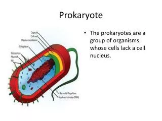
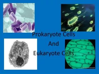
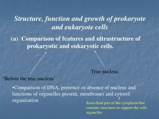
Prokaryote Cells Conclusions
Small Sub Unit 30S 16S RNA 21 proteins Large Subunit 50S 23S & 5S RNAs 31 proteins Bacterial Ribosome
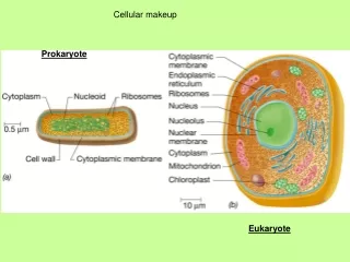
Ribosomes • Complex structures consisting of protein and RNA • Sites of protein synthesis • Smaller than eucaryotic ribosomes • procaryotic ribosomes Þ 70S • eucaryotic ribosomes Þ 80S • S = Svedburg unit
Ribosomal Complexity • Three Dimensional image of the 30s ribosomal subunit • Vital in protein synthesis • Binds to the messenger RNA to initiate translation
50s Ribosomal subunit • The large subunit (50S) from Deinococcus radiodurans contains 33 different proteins • Two rRNA chains (23S and 5S rRNA). The ribosomal rRNA • Responsible for binding t RNA and the catalysis of peptide bonds for translation
16 s Ribosomal subunit from E. coliCulpepper Group at the Stanford School of Medicine
Inclusions • These are storage bodies in the cytoplasm of bacteria • The inclusions vary with the type of bacteria • Provide a supply of vital compounds or ions for metabolism • Reduce osmotic pressure by tying up molecules in particulate form
Inclusions in Cyanobacteria • Cyanophycin granules are found in Cyanobacteria. They are large inclusion bodies composed of polypeptides comprised of arginine and aspartic acid. These store additional nitrogen for the bacteria.
Inclusion bodies • Cyanophycin granules are found in the filamentous photosynthetic bacteria found in fresh water ponds that are vital to the nitrogen cycle in aquatic environments
Carboxysomes • Cyanobacteria, thiobacilli, and nitrifying bacteria, organisms that reduce CO2 in order to produce carbohydrates, possess carboxysomes containing an enzyme used for CO2 fixation. • These may be separated from the cytoplasm by internal membrane
PHB • Poly- hydroxybutyrate molecules joined by ester bonds between the carboxyl and hydroxyl of adjacent molecules. • These are common in purple sulfur bacteria and stain with Sudan black for light microscopy. These granules serve as storage reservoirs for glycogen and sugars necessary for energy and biosynthesis.
Volutin • Some bacteria produce inorganic inclusion bodies in their cytoplasm, including volutin granules that store phosphate and sulfur granules that store sulfur. Volutin is a source of phosphate for DNA. Sulfur is used by purple photosynthetic bacteria that use hydrogen sulfide as a photosynthetic electron donor.
Gas Vacuoles • Purple and green photosynthetic bacteria as well as some other aquatic bacteria contain gas vacuoles. These are aggregates of hollow protein cylinders called gas vesicles that are permeable to atmospheric gas, enabling the organism to regulate buoyancy. Bacteria are able to regulate the depth at which they float to regulate photosynthetic activity
Enterosomes • In Salmonella and E. coli have internal structures similar to carboxysomes • Enterosomes contain enzymes required for the metabolism of certain molecules • The existence of these molecules may be due to the necessity of dealing with toxic molecules • Propanediol is a metabolite of fucose which is a sugar found on the intestinal wall of mammals that that can be degraded by intestinal bacteria – This is one of the molecules metabolized in enterosomes
Magnetosomes • Some motile aquatic bacteria are able to orient themselves by responding to the magnetic fields of the earth because they possess magnetosomes, membrane-bound crystals of magnetite or other iron-containing substances that function as tiny magnets.
External Structures • Fimbriae • Pili • Flagella
Bacterial pili • http://biophysics.bumc.bu.edu/faculty/bullitt/images/cartoon_ppili_hib.jpg
Pili • Pili are appendages that are larger than fimbriae. Their presence is determined by genes on plasmids called sex factors. These structures function in conjugation which is a genetic exchange occurring in bacteria with these appendages
Fimbriae • Fimbriae are thin, hair-lie projections extending from the cell wall in Gram – bacteria. They are composed of helical protein units and designed for attachment to the host cell membranes ( mucous). • They also may contribute to types of movement in some bacteria. • These are considered to be virulence factors and induce many pathogenic effects Neisseria gonorrhea
Fimbriae and Adhesins • The tips of these structures have tips with adhesive proteins called adhesins • They are designed to attach to a specific molecular target • Fimbriae are produced in the cytoplasm and transported to the exterior of the cell
Structural polymorphism of bacterial adhesion pili.Bullitt E, and Makowski L. • Bacterial adhesion pili are designed to bind specifically and maintain attachment of bacteria to target cells. Uropathogenic P-pili are sufficiently mechanically resilient to resist the cleansing action of urine flow that removes most other bacteria. P-pili are 68 A in diameter and approximately 1 micron long, and are composed of approximately 1,000 copies of the principal structural protein, PapA. They are attached to the outer membrane by a minor structural protein, PapH and are terminated by an approximately 20 A diameter fibrillus composed of PapK, PapE and PapF, which presents the host-binding adhesin PapG. The amino-acid sequences of PapA, PapE, and PapF are similar, with highly conserved C-termini being responsible for binding to PapD, the periplasmic chaperone. Our three-dimensional reconstruction indicates that pili are formed by the tight winding of a much thinner structure. A structural transition allows the pilus to unravel without depolymerizing, producing a thin, extended structure five times the length of the original pilus.
Neisseria gonorrhea • To cause infection, Neisseria gonorrhoeae(inf) must first colonize a mucosal surface composed of columnar epithelial cells. Pili alow for this initial binding and, in fact, N. gonorrhoeae is able to rapidly lose pili and synthesize new ones with a different adhesive tip, enabling the bacterium to adhere to a variety of tissues and cells including sperm, the epithelial cells of the mucous membranes lining the throat, genitourinary tract, rectum, and the conjunctiva of the eye. Subsequently, the bacterium is able to make more intimate contact with the host cell surface by way of a cell wall adhesin called Opa
E. Coli and adhesion • http://medschool.umaryland.edu/infeMSD/Images.htm • http://medschool.umaryland.edu/infeMSD/som.html ( Donnenberg lab at University of Maryland)
Flagella Motility http://www-micro.msb.le.ac.uk/video/motility.html
Arrangement of flagella • monotrichous – one flagellum • polar flagellum – flagellum at end of cell • amphitrichous – one flagellum at each end of cell • lophotrichous – cluster of flagella at one or both ends • peritrichous – spread over entire surface of cell
The filament • Hollow, rigid cylinder • Composed of the protein flagellin • Some procaryotes have a sheath around filament
Flagellin( Protein structure) • http://www.rcsb.org/pdb/home/home.do • Search with flagellin • Choose 1ucu • Click on choice • Choose the different image files to learn about molecular structure
References on Genes and Proteins • http://www.ncbi.nlm.nih.gov/ • Choose structures – proteins • Choose nucleotide – genes – DNA sequence • Choose protein – AA sequence • Cn3D – free download to study protein structure
Tubulin subunits of eukaryote flagellum • Tubulin dimer
The three parts of the flagellum • 3 parts • filament • basal body • hook
Hook and Base Structure • http://molvis.sdsc.edu/atlas/morphs/flaghook/index.htm • http://www.umass.edu/microbio/chime/pe_beta/pe/atlas/atlas.htm • http://atlas.proteinexplorer.org
The hook and basal body • Hooklinks filament to basal body • Basal bodyseries of rings that drive flagellar motor
Flagellar Synthesis • An example of self-assembly • Complex process involving many genes and gene products • New molecules of flagellin are transported through the hollow filament • Growth is from tip, not base
Flagellar Motion • flagellum rotates like a propeller • in general, counterclockwise rotation causes forward motion (run) • in general, clockwise rotation disrupts run causing a tumble (twiddle)
Traveling toward and Attractant • Caused by lowering the frequency of tumbles • Traveling away involves similar but opposite responses