Download
1 / 1

E N D
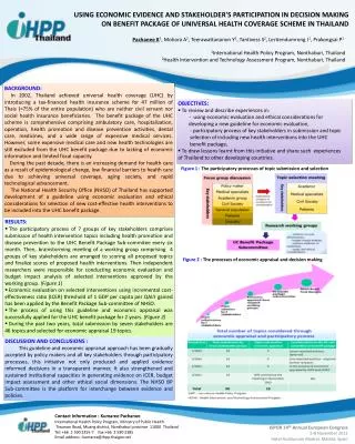
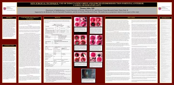
CASE REPORT AND SURGICAL TECHNIQUE RESULTS DISCUSSION On postoperative day 1, there was no ICG in the cornea on biomicroscopic examination using the slit-lamp (Figure 7). The donor cornea was well approximated to the host Descemet’s membrane without Descemet’s membrane folds, air pockets, or debris in the interface between the donor corneal stroma and the host Descemet’s membrane (Figure 7). I introduced a new technique after 100% removal of the host corneal stroma up to the smooth Descemet’s membrane in the area of trephination (7.5 mm), and this space is filled with 100% donor corneal stroma and epithelium after removal of the donor endothelium and Descemet’s membrane. It is imperative to completely expose Descemet’s membrane in the area of trephination, e.g., 7.5 mm diameter. TALK does not apply to dissections in which Descemet’s membrane is only partially exposed in the visual axis centrally,2,6 while host corneal stroma Descemet’s membrane is in the periphery. TALK provides a more natural posterior corneal curvature and excellent smooth apposition of the host Descemet’s membrane to the donor corneal stroma without folds or air pockets at the donor (stroma)-host (Descemet’s membrane) interface (Figure 7). In contrast, other ALK procedures in which varying amounts of residual host corneal stroma and an oversized donor corneal disk (0.25 to 0.5 mm oversize) are used3 may alter the posterior corneal curvature and possibly contribute to the formation of host Descemet’s membrane folds postoperatively. Use of a corneal diameter that is the same size as the host corneal trephination also helps prevent postoperative Descemet’s membrane folds. In addition, removal of 100% host corneal stroma followed by grafting of the donor corneal stroma of the same diameter, also 100% thickness, helps to even out the host-graft junction on the corneal epithelial surface without the donor tissue being elevated above the surrounding peripheral host cornea. Classification of ALK We propose a new classification for ALK as shown in Table 1. To date, the term ALK is applied to any lamellar dissection in which Descemet’s membrane and host endothelium are preserved. This encompasses a wide range of corneal depth for the lamellar dissection with varying quality of the optical interface. The proposed classification is primarily based on the depth of the host corneal lamellar dissection. Depending on the depth of the corneal dissection, the level of the corneal interface between the donor and host cornea varies in the operated cornea. In superficial lamellar keratoplasty (SLK), the interface is at 30% or less, while in TALK the interface is at 100% thickness at the level of the host Descemet’s membrane (Table 1). This classification, therefore, provides a better understanding of the interface location within the host cornea and its role in the final visual acuity postoperatively. The deeper the donor-host interface within the transplanted cornea, the clearer the interface should be, which may result in earlier visual rehabilitation. Future studies will prove or disprove this theory. In fact, Barraquer recommended deeper dissections for lamellar keratoplasty.46If this hypothesis is correct, TALK may facilitate earlier and possibly better visual outcomes compared to SLK, mid-lamellar keratoplasty (MLK), or posterior lamellar keratoplasty (PLK). Terminology Descriptions of lamellar corneal surgery can be confusing with various terminologies have been used in the literature, including lamellar keratoplasty (LK),1 deep lamellar anterior keratoplasty (DLAK),1 DLEK,1 deep stromal anterior lamellar keratoplasty (DSALK),3 deep lamellar keratoplasty (DLKP),18 DLK,2 deep anterior lamellar keratoplasty (DALK),26 and PLK.27 I propose the following unified definitions for lamellar keratoplasty: 1) anterior lamellar keratoplasty (ALK) and 2) posterior lamellar keratoplasty (PLK). In this manuscript, ALK is defined as any corneal lamellar procedure in which the host Descemet’s membrane and endothelium are retained, and PLK is defined as any corneal lamellar procedure in which Descemet’s membrane and the endothelium are excised with or without host corneal stroma. ALK would include SLK, MLK, DLK, and TALK (Table 1) and PLK would include DLEK and transplantation of Descemet’s membrane and endothelium.47 Unlike ALK, in PK there is no interface in the visual axis of the patient’s cornea, which provides maximum optical clarity. The only interface in PK is at the host-graft junction 3600, at varying distances from the anatomic center of the cornea, depending on the diameter of the host corneal trephination. In contrast, in ALK, there is an interface in the region of the visual axis and at the host-graft junction 3600. We define the corneal interface as the plane of physical contact between the donor and host corneas. This interface in ALK is like a filter that is maximally opaque in the immediate postoperative period and that increases in clarity over time, until this filter effect of the corneal interface completely disappears over time, which in my experience may take as long as 1 year. I have also found that this corneal interface in ALK is more opaque when the interface is stroma-to-stroma, as in SLK, MLK, and DLK, and this interface is least dense between the stroma and Descemet’s membrane, as in TALK. Future studies may determine whether TALK provides earlier visual rehabilitation compared to SLK, MLK, or DLK. The clarity of the corneal interface usually plays a role in the best-corrected visual acuity. The difficulty with deeper lamellar dissection is the completion of the dissection without perforating the AC. To facilitate deep lamellar dissection, various techniques including air injection,40-43 fluid injection,44 viscoelastic injection,45 and a combination of intracorneal viscoelastic and air in the AC3,26have been used previously. However, each has limitations. In addition, various complications have been associated with these techniques including, a fixed, dilated pupil (Urrets-Zavalia syndrome) after the use of air/gas in the AC in DLK,48 possible endothelial cell damage secondary to air in the AC during surgery,49,50 and sequestration of viscoelastic in the cornea causing substantial patient morbidity that requires PK following viscoelastic delamination in DLK.51 Sometimes, the viscoelastic may be present in the corneal interface even 1 month after ALK and requires another operation to remove it.3 In addition, in ALK with a scleral approach (Table 2), Viscoat is injected via the scleral incision into the corneal stromal interface to create an artificial AC,3 and in so doing, Descemet’s membrane is deformed from its resting convex surface to an altered concave surface. At the end of ALK, folds can develop if Descemet’s membrane has not fully reverted back to its normal convex configuration. In TALK, there is no deformation of the resting convex state of the host Descemet’s membrane and therefore less chance of iatrogenic induction of folds at the end of TALK. Other differences between the scleral and the corneal approach in ALK are listed in Table 2. The formation of the whitish-green color secondary to ICG and forced hydrodissection facilitates viewing of the split corneal stroma and enhances the lamellar dissection and raises the surgeon comfort level regarding preserving the AC. Using this technique of ICG with forced hydrodissection, the surgeon can fully excise the corneal stroma (100% corneal stromal thickness) and expose the smooth Descemet’s membrane surface in the entire trephination area (diameter 7.5 mm). This ICG enhanced corneal stromal dissection eliminates the need for air injection into the AC to assess the stromal thickness from Descemet’s membrane. In addition, unlike other techniques in which only the central region of Descemet’s membrane is exposed,2,6 in TALK, the entire Descemet’s membrane within the diameter of trephination (7.5 mm) is exposed without perforating the AC. This layer-by-layer stromal dissection should be carried out slowly, with time taken to achieve the final goal of fully exposing Descemet’s membrane within the 7.5 mm diameter and not perforating it. Merocel eye spears are used in TALK (as in LASIK) to prevent introduction of debris between the donor corneal stroma and the host Descemet’s membrane. For the same reason, excess irrigation is carried out on the donor stromal surface and the host Descemet’s membrane before placing the donor disk over the host Descemet’s membrane and suturing it in place. Barraquer outlined the conditions favorable for obtaining better visual results with LK including the notion of the deepest possible interface to reduce scarring and performing smooth surface sectioning of the graft and the bed.46 The corneal intrastromal interface, as in SLK, MLK, and DLK, has keratocytes and other tissue elements on either side of the interface, which may play a role in the continued remodeling of the corneal interface. In TALK, there are keratocytes only on the stromal side of the stromal-Descemet’s membrane interface. In addition, in manual surgical dissections in SLK, MLK, and DLK, there are irregularities in the corneal intrastromal interface. In TALK, at least 50% of the interface is smooth since the exposed Descemet’s membrane has no corneal stromal fibers and a smooth surface (Figure 6,A). In addition, the donor corneal stroma is expected to be at least 90% or more smooth, since the surgical steps only involve peeling of Descemet’s membrane with its attached endothelium and no lamellar dissection is performed (Figure 6,C). Thus, the increased smoothness of the stromal-Descemet’s membrane interface and the 100% depth of the interface in TALK may contribute to increased clarity of the interface compared to other manually created intrastromal interfaces, whether it is SLK, MLK, or DLK. If so, TALK may become the preferred choice of all ALK procedures over time. In TALK there is no host corneal stromal bed, rather only Descemet’s membrane with its endothelium. The question arises if these patients are susceptible to corneal ectasia since there is no stromal bed. I do not expect corneal ectasia in TALK, since the donor corneal thickness is 100%, except for Descemet’s membrane and its endothelium, thus preserving the host total corneal thickness. The epithelial surface of the donor-host junction is at the same level and 10-0 nylon sutures are used similar to PK. In addition, in PK corneal ectasia usually does not occur. Although automated microkeratomes have resulted in major improvements in technology,1 it is impossible to use them at the present time for TALK, because Descemet’s membrane would be perforated and the AC entered. Recently, the femtosecond laser FEMTEC (20/10 Perfect Vision, Heidelberg, Germany) was used in PLK in enucleated porcine eyes and human donor corneas.52 The cut edges had an electron-dense layer as seen by TEM.52 Whether laser technology can recognize and specifically treat the host stromal- Descemet’s membrane interface without tearing it or leaving behind stromal fibers attached to it has not been determined. The stromal-Descemet’s membrane interface is a specific plane that is convex, and at present only manual dissection appears to successfully cleave this interface and result in a smooth DM without residual corneal stromal fibers. Presently, laser technology and automated microkeratomes cannot be used to successfully perform TALK. This report pertains to optical lamellar keratoplasty and not tectonic lamellar keratoplasty,53-62which may be used for various ocular indications including corneal melt and corneal perforation. The prevailing technique of air injection into the AC provides an air-to-endothelial interface in ALK,3 but even with this interface it is difficult to remove all stroma overlying Descemet’s membrane without perforating the AC. Air injection into the AC combined with intrastromal viscoelastic has been associated with perforation into the AC in 12% of cases at 95% average corneal depth3 and perforations in 33% of cases with deeper dissections, i.e., 99% corneal depth was attempted with air in the AC and no intrastromal viscoelastic.63 In a larger series of 120 human eyes undergoing optical DLK for visual improvement, the perforation rate was 39%.2 This rate may be considered high when deep corneal dissections are attempted. When intrastromal air was used for DLK, the intraoperative perforation rate was 30% and resulted in conversion to PK in these cases.41 Chau et al.42 evaluated air LK and found that air dissection was relatively incomplete, leaving the region with the most stromal scarring or pathology undissected. Thus, until the present report, there has been no ideal surgical technique in which TALK could be performed without perforation and effective removal of all stromal fibers to fully expose the bare Descemet’s membrane in the area of trephination. The use of ICG with forced hydrodissection appears to be superior to air injection into the AC, and it may become the preferred surgical method for TALK. In addition, injection of air into the AC converts TALK from an extraocular to an intraocular procedure with possible complications including infection, endophthalmitis, and possible damage to the intraocular structures. With the use of ICG and forced hydrodissection, TALK remains an extraocular procedure and hence may be safer compared to lamellar surgery with air injection into the AC.3 ICG stains any exposed stroma and because of this the surgeon does not have to rely on intraocular air injection. ICG is a sterile, water-soluble, tricarbocyanine dye with a peak spectral absorption of 800 to 810 nm in blood plasma or blood.64 ICG is taken up from the plasma almost exclusively by the hepatic parenchymal cells and secreted entirely into the bile. 64 ICG contains not more than 5.0% sodium iodide.64 ICG is marketed for intravenous administration. Anaphylactic or urticarial reactions were reported in patients with or without a history of allergy to iodides.64 If reactions occur, treatment with appropriate agents, e.g., epinephrine, antihistamines, and corticosteroids, should be initiated. It is unknown whether this drug is excreted in human milk,64 and caution should be exercised when ICG is administered to a nursing woman.64 ICG has been widely used in various medical fields, including cardiovascular, hepatic, and ophthalmic specialties,65 and used extensively in animal66-73 and human studies.74-78 In ophthalmology, it was used initially for ophthalmic angiographic studies and more recently to stain the anterior lens capsule under air before capsulorrhexis during phacoemulsification and in DLEK.7 The effect of ICG on donor keratocytes, if any, has not been evaluated. However, in TALK there is no direct exposure of stromal keratocytes to ICG on the donor corneal disk and all stromal fibers are excised in the host corneal dissection. When ICG is used in the AC under air, the aqueous humor in the AC immediately dilutes the injected dye. Because no dilution occurs when the dye is added directly to the exposed host corneal stroma and the donor corneal endothelium, the dye is diluted further 1:1 with sterile BSS before its application to the host corneal stroma and donor corneal endothelium in TALK. Following host corneal exposure to this dye, ICG selectively stains only the exposed donor corneal stroma and not the uncut, full-thickness, host corneal rim with intact epithelium. There is never direct introduction of ICG intraocularly in TALK. There are several advantages to the intraoperative use of ICG dye to stain the donor corneal stroma (Table 3). ICG in combination with forced hydrodissection greatly facilitates performing this new surgical procedure and contributes to a good postoperative result, with no ICG seen the day after surgery (Figure 7). This is the first description of the combined use of ICG and forced hydrodissection in TALK, which eliminates the use of intraocular air and maintains TALK as a completely extraocular procedure. It is also the first use of ICG to stain the donor corneal endothelium. The major surgical steps of TALK are shown in the intraoperative photographs (Figures 1-6). Figure 7 shows the view on day 1 postoperatively. Case 1 is a 50-year-old woman with advanced keratoconus (Figure 1,A) and contact lens failure who underwent TALK (7.5 mm/7.5 mm) in her right eye. Case 2 is a 76-year-old man with an anterior corneal scar secondary to an infectious bacterial corneal ulcer who underwent TALK (7.5 mm/7.5 mm) in his left eye. ICG Preparation ICG (IC-Green, Akorn, Inc., Buffalo Grove, IL) is packaged in a 25-mg vial and a 10-ml ampule of aqueous solvent (Akorn, Inc.). The aqueous solvent (pH 5.5-6.5) is prepared sterile water for injection used to dissolve the ICG. Aqueous solvent, 0.5 ml, is added to the ICG bottle and 4.5 ml of sterile BSS is added to the bottle containing the ICG. The bottle is shaken until the contents are well dissolved. Two ml of the ICG solution is aspirated into a 5-ml sterile syringe and another 2 ml of sterile BSS is aspirated into the same syringe for a total volume of 4 cc (1:1 dilution). A sterile, blunt, 30-gauge cannula is attached to the syringe and the solution is ready for corneal use as described below. Host Corneal Surgery with ICG and Hydrodissection The central cornea was marked after being measured with Castroviejo calipers. A Barron radial vacuum trephine (Katena Products, Inc., Denville, NJ) 7.5 mm in diameter was centered over the corneal mark using the built-in cross wires for proper centration. Suction is applied to the trephine and partial-thickness trephination is carried out (about 70% corneal thickness) (Figure 1,B). The corneal markings help in proper suture placement (Figure 1,C). The plane of stromal dissection is initiated using a Crescent knife (BeaverTM, Becton Dickinson & Co., Franklin Lakes, NJ), angled 550, bevel-up, matte finish and then switched to a Morlet lamellar knife/dissector (#6-607, Duckworth & Kent USA, Ltd., St. Louis, MO) (Figure 1,D). The lamellar corneal dissector (long arm of the dissector) is used to dissect off the host corneal cap, which is about 70% corneal thickness from the anterior corneal surface. This exposes the remaining host corneal stromal bed (about 30% corneal thickness) (Figure 2,A). A 150 super-blade (Alcon ophthalmic knife 150, Alcon Surgical, Fort Worth, Texas) or the angled Crescent knife then is used to make a partial cut into the remaining corneal stroma, with care taken not to perforate Descemet’s membrane. This stromal opening is used to place a smooth 30-gauge AC cannula (Alcon Surgical) with the opening of the cannula facing down, and BSS is injected under pressure using a 5-cc disposable syringe. This forced hydrodissection further splits the remaining corneal stromal layers, and the superficial layer of the corneal stroma turns white in the immediate segmental area (Figure 2,B). ICG is used to fill the well created in the host cornea with the trephination and dissecting off of the corneal cap. The excess ICG is removed immediately using sterile Merocel eye spears (Medtronic Solan, Jacksonville, FL). The exposed host corneal stroma is green, which facilitates further layer-by-layer stromal dissection and the segmental area of hydrodissection turns whitish-green. The short arm of the Morlet lamellar knife/dissector, i.e., the Paufique knife, then is introduced gently into this focal pocket and intrastromal lamellar dissection is carried out (Figure 2,C) by a to-and-fro 1800 arc rotation on the vertical axis of the handle of the Morlet lamellar knife/dissector held perpendicular to the stromal bed. Following segmental lamellar dissection of the corneal stroma, a Vannas scissor blade is introduced gently into the dissected pocket and cut vertically while the anterior stromal layer is simultaneously lifted away from the remaining stromal bed (Figure 3,A). This segmentally separated green stromal layer exposes in sharp contrast the relatively transparent, unstained deeper stromal layers (Figure 3,B). The separated, green stained, segmental stromal layer then is excised with the Vannas scissors. This creates a transparent area in the region of the excised stromal layer, surrounded by green stroma. This segmental hydrodissection and layered excision of the corneal stromal bed is repeated (Figure 3,C, D, Figure 4,A, B, C, D) until all green stroma is excised and the entire 7.5-mm circular area is transparent. The well is filled with ICG (Figure 5, A), and the excess ICG is removed using Merocel eye spears (Figure 5,B). ICG stains the corneal stroma instantaneously. A vertical cut is again made using a 150 super-blade or the angled Crescent knife on the remaining thin corneal stroma and hydrodissection and layer-by-layer segmental excision of the remaining corneal stroma is performed (Figure 5,C) until once again the 7.5 mm diameter is fully transparent. After this, a final layer of stroma is removed similarly (Figure 5,D) exposing the smooth Descemet’s membrane with no corneal stroma in the entire area of the 7.5 mm diameter (Figure 6,A). There is a clearly demarcated light reflex on the smooth Descemet’s membrane (Figure 6,A). If there are stromal fibers on the surface of the Descemet’s membrane, the light reflex will be fragmented. The presence of a clear, sharp light reflex on Descemet’s membrane confirms visually that the bare Descemet’s membrane has been reached with no stromal fibers. If there is residual stroma after the third layer is removed, this dissection process can be repeated until bare Descemet’s membrane is exposed in the 7.5-mm diameter area without corneal stroma. Donor Corneal Surgery with ICG Staining of Endothelium The donor corneal button is placed on a Teflon block with the endothelial side up, and the concavity in the corneal button is filled with ICG (Figure 6,B). Excess ICG is removed with Merocel eye spears. Vacuum is applied to the epithelial side of the donor cornea and trephination is carried out using a disposable 7.5-mm Hanna trephine blade (Moria, Antony, France; distributed by Microtech, Inc., Doylestown, PA). The scleral rim with the attached peripheral donor cornea is removed. Vacuum is applied again to the epithelial side of the donor disc 7.5 mm diameter and the donor Descemet’s membrane with the green endothelium (Figure 6,C) is peeled from the donor corneal stroma, which exposes the donor corneal stroma without any ICG. Donor Disk Transplantation The interface is rinsed thoroughly with sterile BSS, i.e., the stromal side of the donor disk and the host Descemet’s membrane. The donor corneal disk then is placed over the smooth host Descemet’s membrane and sutured in place with 10-0 nylon interrupted and a running 3600 suture (Figure 6,D). Suture knots are buried within the host stroma. Astigmatism is controlled by intraoperative suture adjustments as needed. A collagen corneal shield (ProshiledTM RD, Alcon Laboratories) soaked in TobraDex is placed over the cornea. Table 1.Classification of ALK. A. Based on the corneal depth of dissection Terminology Depth Interface Risk of AC entry Superficial lamellar keratoplasty 30% Stroma-to-stromaNo (SLK) Mid-lamellar keratoplasty (MLK)30-70%Stroma-to-stromaNo Deep lamellar keratoplasty (DLK)70-95%Stroma-to-stromaYes Total lamellar keratoplasty (TALK)100%Stroma-to-DMYes DM – Descemet’s membrane Figure 2. A: Exposed host corneal stromal bed of about 30% corneal thickness after excision of the host corneal cap of about 70% corneal thickness. The cornea is relatively transparent. B: A 30-G smooth cannula is placed in the pocket created by the vertical cut using a 150 super-blade, with the cannula opening directed toward Descemet’s membrane; forced hydrodissection using BSS creates the whitish opacity of the split anterior layer of the corneal stroma in a segmental area. C: The corneal well, 7.5 mm in diameter, is filled with ICG. D: The Paufique knife (short arm of the Morlet lamellar knife/dissector) is introduced into the focal pocket created by the forced hydrodissection. The green stromal layer anterior to the Paufique knife is easily seen and facilitates dissection. Figure 1.A: Intraoperative photograph shows keratoconus. B: Barron radial vacuum trephine is held in place on the ocular surface with suction and partial-thickness (about 70% corneal thickness) trephination is carried out on the host cornea. C: Corneal marks on the host cornea following partial-thickness trephination allow for accurate suture placement. D: Morlet lamellar knife/dissector (long arm of the instrument) is used to dissect off the host corneal cap of about 70% corneal thickness from the corneal surface. B. Based on the indication for surgery Indication Type of Lamellar Keratoplasty Therapeutic purposeTectonic lamellar keratoplasty For visual purposeOptical lamellar keratoplasty: SLK, MLK, DLK, TALK C. Based on surgical approach Surgical Approach Route of Entry for Corneal Lamellar Dissection DirectCornea IndirectScleral-pocket INTRODUCTION Figure 3.A: The focal pocket that is dissected is lifted and cut with a Vannas scissor.B: Separating the flaps of this pocket reveals the transparent corneal stroma that is remaining, in sharp contrast against the green ICG stained more superficial corneal stroma that is reflected. C: Additional forced hydrodissection with BSS is carried out on either side of the excised corneal stromal flap. The stromal layer that is hydrodissected appears whitish green in sharp contrast to the transparent corneal stroma in between these two islands. D: Further forced hydrodissection with BSS is carried out. Figure 4. A: Forced hydrodissection of the peripheral corneal stroma close to the trephination boundary is carried out. The ICG green stain highlights the remaining layer of stroma in that corneal plane that is not yet removed. The transparent area where the ICG stained corneal layer at the same plane that has already been removed. B: ICG stained corneal stromal layer close to the trephination boundary being excised with Vannas scissors.C: Paufique knife being introduced between the ICG stained corneal stromal layer in the far periphery close to the trephination mark and the remaining corneal stromal bed. D: Corneal layer being excised with Vannas scissors. Table 2.Comparison of indirect scleral-pocket approach to direct corneal approach in anterior lamellar keratoplasty. Description Scleral-pocket Cornea Approach with ICG Type of ocular procedureConverts an extraocular Fully extraocular procedure procedure to an intraocular procedure by air injection into AC ViscoelasticRequiredNot required Intrastromal viscoelastic Possible Not applicable retention and second surgery Additional scleral wound Yes No with possible added astigmatism Graft sizeOversize graft with possible Same size graft with no DM folds DM folds Depth assessment of lamellar Relies on air-to-endothelial Direct view of ICG-stained Dissection light reflex corneal stroma Dissection depth90 – 95%100% Full-thickness donor cornea YesYes without endothelium and DM DM – Descemet’s membrane Lamellar corneal surgery, both anterior lamellar keratoplasty (ALK) and posterior lamellar keratoplasty (PLK), recently have gained attention among anterior segment surgeons and cornea specialists1-39 largely because of various improvements in the surgical steps and instrumentation. Preservation of the host corneal surface and anterior cornea, as in deep lamellar endothelial keratoplasty (DLEK), or preservation of the host corneal endothelium and Descemet’s membrane, as in ALK, has potential advantages for the host. In ALK, graft rejection can be prevented, which is advantageous to the patient. However, these lamellar surgical procedures are challenging, because the cornea has to be split by lamellar dissection and not perforated either anteriorly onto the corneal surface or posteriorly through Descemet’s membrane into the anterior chamber (AC). Any substantial perforation results in termination of the lamellar surgery and conversion to the backup surgical procedure, usually a full-thickness penetrating keratoplasty (PK). Previous authors have described the use of intracorneal air injection,40-43 fluid injection,44 viscoelastic injection,45 and a combination of intracorneal viscoelastic and air in the AC3,26 to split the corneal stroma in a single plane. Air, viscoelastic, and fluid hydrodissection have limitations in its usefulness in ALK, and air in the AC makes ALK an intraocular procedure with potential complications. I previously described the use of indocyanine green (ICG) in DLEK.7 In the present report, I describe the combined use of ICG and forced hydrodissection using balanced salt solution (BSS), which greatly facilitates the layer-by-layer dissection of the host corneal stroma to fully expose the host Descemet’s membrane to the full diameter of the trephination mark, unlike previous descriptions in which Descemet’s membrane is to be exposed only in the central optical area.2,6 ICG is also used to stain the donor endothelium before it is removed along with the attached donor Descemet’s membrane from the remaining donor corneal stroma. The entire donor corneal stroma is transplanted onto the fully exposed smooth host Descemet’s membrane; hence, this procedure is called total anterior lamellar keratoplasty (TALK). This is the first use of ICG in TALK both in the host cornea and on the donor corneal endothelium. Figure 6. A: Descemet’s membrane is fully exposed in the area of the 7.5-mm diameter. The sharp margins of the light reflex, confirming the end point of the surgical dissection, namely, the smooth DM without stromal fibers. B: ICG fills the concavity of the donor corneal button on the endothelial side. C: ICG-stained donor corneal endothelium and its attached Descemet’s membrane are peeled from the donor corneal stroma. D: Donor corneal stroma is placed on the bare Descemet’s membrane and sutured in place with 10-nylon interrupted and running sutures with buried knots. Figure 5. A: ICG filled corneal well to stain the remaining corneal stromal layer close to Descemet’s membrane.B: Excess ICG is removed using Merocel eye spear. C:Paufique knife dissection of the remaining ICG-stained thin corneal stroma.D:Further Paufique knife dissection of the remaining ICG-stained thin corneal stroma. The segmental area of the bare Descemet’s membrane is exposed. Table 3.Advantages of intraoperative use of ICG with forced hydrodissection in TALK. 1. The host corneal stromal tissue stains instantly from clear to green 2. Forced hydrodissection beneath the green stained stroma makes it whitish-green and greatly facilitates layer-by-layer stromal dissection 3. Staining takes less than 60 seconds with no significant surgical delay 4. ICG eliminates the need for air injection into the AC and keeps TALK a fully extraocular procedure 5. ICG disappears from the host cornea within 24 hours after surgery 6. ICG with forced hydrodissection makes it possible to fully excise the host corneal stroma 100%, and fully expose the smooth host DM in the entire area of trephination 7. ICG stains the donor endothelium and facilitates visualization of the donor endothelium- DM complex for its surgical removal leaving behind 100% donor corneal stroma and the surface epithelium for transplantation DM—Descemet’s membrane. Figure 7. Slit-lamp photograph on postoperative day 1 shows excellent apposition of the donor corneal stroma (100% thickness) to the smooth host Descemet’s membrane (DM) without folds in the membrane, interface air pockets, or debris. No ICG is seen in the cornea. Continuous and interrupted 10-0 nylon sutures are visible. The epithelial surface of the donor-host junction is even without vertical size disparity. NEW SURGICAL TECHNIQUE: USE OF INDOCYANINE GREEN AND FORCED HYDRODISSECTION FORTOTAL ANTERIOR LAMELLAR KERATOPLASTY Thomas John, MD Department of Ophthalmology, Loyola University at Chicago, Maywood, IL; and Chicago Cornea Research Center, Tinley Park, IL Supported by the Richard A. Perritt Charitable Foundation, and the Loyola Cornea Research Fund.The author has no proprietary interest in any aspect of this study. ABSTRACT REFERENCES 1. Terry MA. The evolution of lamellar grafting techniques over twenty-five years. Cornea 2000; 19:611-616 2. Sugita J, Kondo J. Deep lamellar keratoplasty with complete removal of pathological stroma for vision improvement. Br J Ophthalmol 1997; 81:184-188 3. Melles GRJ, Lander F, Rietveld FJ, et al. A new surgical technique for deep stromal, anterior lamellar keratoplasty. Br J Ophthalmol 1999; 83:327-333 4. Shimazaki J. The evolution of lamellar keratoplasty. Curr Opin Ophthalmol 2000; 11:217-223 5. Rich LF. Expanding the scope of lamellar keratoplasty. Trans Am Ophthalmol Soc 1999; 97:771-814 6. Tsubota K, Kaido M, Monden Y, et al. A new surgical technique for deep lamellar keratoplasty with single running suture adjustment. Am J Ophthalmol 1998; 126:1-8 7. John T. Use of indocyanine green in deep lamellar endothelial keratoplasty. J Cataract Refract Surg 2003; 29:437-443 8. Polack FM. Lamellar keratoplasty. Malbran’s “peeling off” technique. Arch Ophthalmol 1971; 86:293-295 9. Panda A, Bageshwar LM, Ray M, et al. Deep lamellar keratoplasty versus penetrating keratoplasty for corneal lesions. Cornea 1999; 18:172-175 10. Jain S, Azar DT. New lamellar keratoplasty techniques: posterior keratoplasty and deep lamellar keratoplasty. Curr Opin Ophthalmol 2001; 12:262-268 11. Barraquer JI. Lamellar keratoplasty (special techniques). Ann Ophthalmol 1972; 4:437-469 12. Terry MA, Ousley PJ. Endothelial replacement without surface corneal incisions or sutures; topography of the deep lamellar endothelial keratoplasty procedure. Cornea 2001; 20:14-18 13. Gasset AR. Lamellar keratoplasty in the treatment of keratoconus: conectomy. Ophthalmol Surg 1979; 10:26-33 14. Melles GR, Lander F, Nieuwendal C. Sutureless posterior lamellar keratoplasty: a case report of a modified technique. Cornea 2002; 21:325-327 15. Trimarchi F, Poppi E, Klersy C, Piacentini C. Deep lamellar keratoplasty. Ophthalmologica 2001; 215:389-393 16. Terry MA, Ousley PJ. Deep lamellar endothelial keratoplasty in the first United States patients; early clinical results. Cornea 2001; 20:239-243 17. Anwar M. Dissection technique in lamellar keratoplasty. Br J Ophthalmol 1972; 56:711-713 18. Shimazaki J, Shimmura S, Ishioka M, Tsubota K. Randomized clinical trial of deep lamellar keratoplasty vs penetrating keratoplasty. Am J Ophthalmol 2002; 134:159-165 19. Guell JL, Velasco F, Guerrero E, et al. Preliminary results with posterior lamellar keratoplasty for endothelial failure. Br J Ophthalmol 2003; 87:241-243 20. Saini JS, Jain AK, Sukhija J, Saroha V. Indications and outcome of optical partial thickness lamellar keratoplasty. Cornea 2003; 22:111-113 21. Behrens A, Ellis K, Li L, et al. Endothelial lamellar keratoplasty using an artificial anterior chamber and a microkeratome. Arch Ophthalmol 2003; 121:503-508 22. Melles GR, Eggnik FA, Lander F, et al. A surgical technique for posterior lamellar keratoplasty. Cornea 1998; 17:618-626 23. Melles GRJ, Landers F, van Dooren BTH, et al. Preliminary clinical results of posterior lamellar keratoplasty through a sclerocorneal pocket incision. Ophthalmology 2000; 107:1850-1856 24. Melles GR, Remeijer L, Geerards AJ, Beekhuis WH. The future of lamellar keratoplasty. Curr Opin Ophthalmol 1999; 10:253-259 25. Azar DT, Jain S, Sambursky R. A new surgical technique of microkeratome-assisted deep lamellar keratoplasty with a hinged flap. Arch Ophthalmol 2000; 118:1112-1115 26. Melles GR, Remeijer L, Geerards AJ, Beekhuis WH. A quick surgical technique for deep, anterior lamellar keratoplasty using visco-dissection. Cornea 2000; 19:427-432 27. Seitz B, Langenbucher A, Hofmann-Rummelt C, et al. Nonmechanical posterior lamellar keratoplasty using the femtosecond laser (femto-plak) for corneal endothelial decompensation. Am J Ophthalmol 2003; 136:769-772 28. Melles GR, Lander F, Rietveld FJ, et al. A new surgical technique for deep stromal, anterior lamellar keratoplasty. Br J Ophthalmol 1999; 83:327-333 29. Ehrlich MI, Phinney RB, Mondino BJ, Pettit TH. Techniques of lamellar keratoplasty. Int Ophthalmol Clin 1988; 28:24-29 30. Panda A, Singh R. Intralamellar dissection techniques in lamellar keratoplasty. Cornea 2000; 19:22-25 31. Bourne RA. Visual improvement with lamellar keratoplasty. Can J Ophthalmol 1968; 3:43-45 32. Fasco A, Nagy A, Balazs K, Berta A. Changing techniques and indications for 13 33. 12lamellar keratoplasty. Acta Chir Hung 1997; 36:79-80 34. Soliman MM, el-Din AA, Ayoub MI. Lamellar keratoplasty. Bull Ophthalmol Soc Egypt 1972; 65:103-113 35. Morrison JC, Swan KC. Full-thickness lamellar keratoplasty. A histologic study in human eyes. Ophthalmology 1982; 89:715-719 36. Clayton T, Miller D. Lamellar keratoplasty using a freezing platform. Ophthalmic Surg 1984; 15:592-595 37. Bessant DAR, Dart JKG. Lamellar keratoplasty in the management of inflammatory corneal ulceration and perforation. Eye 1994; 8:22-28 38. Vancea PP, Schwartzenberg T. Lamellar keratoplasty in keratoconus. Albrecht Von Graefes Arch Klin Exp Ophthalmol 1979; 212:55-64 39. McDonnell PJ, Falcon MG. The lamellar corneal graft for optical indications. Eye 1988; 2:390-394 40. Archila EA. Deep lamellar keratoplasty dissection of host tissue with intrastromal air injection. Cornea 1984; 3:217-218 41. Price FW Jr. Air lamellar keratoplasty. Refract Corneal Surg 1989; 5:240-243 42. Chau GK, Dilly SA, Sheard CE, Rostron CK. Deep lamellar keratoplasty on air with lyophilised tissue. Br J Ophthalmol 1992; 76:646-650 43. Anwar M, Teichmann KD. Deep lamellar keratoplasty: surgical techniques for anterior lamellar keratoplasty with and without baring of Descemet’s membrane. Cornea 2002; 21:374-383 44. Amayen AF, Anwar M. Fluid lamellar keratoplasty in keratoconus. Ophthalmology 2000; 107:76-79 45. Manche EE, Holland GN, Maloney RK. Deep lamellar keratoplasty using viscoelastic dissection. Arch Ophthalmol 1999; 117:1561-1565 46. Barraquer J. Lamellar keratoplasty, (special techniques). Ann Ophthalmol 1972; 4:437-469 47. Melles GR, Lander F, Rietveld FJ. Transplantation of Descemet’s membrane carrying viable endothelium through a small scleral incision. Cornea 2002; 21:415-418 48. Maurino V, Allan BD, Stevens JD, Tuft SJ. Fixed dilated pupil (Urrets-Zavalia syndrome) after air/gas injection after deep lamellar keratoplasty for keratoconus. Am J Ophthalmol 2002; 133:266-268 49. Kim EK, Cristol SM, Geroski DH, et al. Corneal endothelial damage by air bubbles during phacoemulsification. Arch Ophthalmol 1997; 115:81-88 50. Eiferman RA, Wilkins EL. The effect of air on human corneal endothelium. Am J Ophthalmol 1981; 92:328-331 51. Bhojwani RD, Noble B, Chakrabarty AK, Stewart OG. Sequestered viscoelastic after deep lamellar keratoplasty using viscodissection. Cornea 2003; 22:371-373 52. Seitz B, Langenbucher A, Hofmann-Rummelt C, et al. Nonmechanical posterior lamellar keratoplasty using the femtosecond laser (femto-plak) foe corneal endothelial decompensation. Am J Ophthalmol 2003; 136:769-772 53. Shimmura S, Shimazaki J, Tsubota K. Therapeutic deep lamellar keratoplasty for corneal perforation. Am J Ophthalmol 2003; 135:896-897 54. Ti SE, Tan DT. Tectonic corneal lamellar grafting for severe scleral melting after pterygium surgery. Ophthalmology 2003; 110:1126-1136 55. Cheng CL, Tan DT. Lamellar corneal autograft for corneal perforation. Aust N Z J Ophthalmol 1999; 27:437-439 56. Titiyal JS, Ray M, Sharma N, et al. Intralamellar autopatch with lamellar keratoplasty for paracentral corneal perforations. Cornea 2002; 21:615-618 57. Hahn TW, Kim JH. Two-step annular tectonic lamellar keratoplasty in severe Terrien’s marginal degeneration. Ophthalmic Surg 1993; 24:831-834 58. Watts P, Michaeli-Cohen A, Abdolell M, Rootman D. Outcome of lamellar keratoplasty for limbal dermoids in children. J AAPOS 2002; 6:209-215 59. Vajpayee RB, Bhartiya P, Sharma N. Central lamellar keratoplasty with peripheral intralamellar tuck: a new surgical technique for keratoglobus. Cornea 2002; 21:657-660 60. Zhou S, Chen J, Fung B, Chen L. Fresh amniotic membrane transplantation combined lamellar keratoplasty for patients with recurrent Mooren’s ulcer. Yan Ke Xue Bao 2003; 19:1-5 61. Jones DH, Kirkness CM. A new surgical technique for keratoglobus-tectonic lamellar keratoplasty followed by secondary penetrating keratoplasty. Cornea 2001; 20:885-887 62. Sanyo Y, Okamoto S, Nishida K, et al. Peripheral lamellar keratoplasty for corneoscleral cyst: three case reports. Cornea 1999; 18:233-236 63. Melles GRJ, Rietveld FRJ, Beekhuis WH, Binder PS. A technique to visualize corneal incision and lamellar dissection depth during surgery. Cornea 1999; 18:80-86 64. Physicians Desk Reference for Ophthalmic Medicines, 30th ed. Montvale, NJ, Medical Economics, 2002; 203-204 65. Maarek JM, Holschneider DP, Harimoto J. Fluorescence of indocyanine green in blood: intensity dependence on concentration and stabilization with sodium polyasparate. J Photochem Photobiol B 2001;65: 157-164 66. Springett R, Sakata Y, Delpy DT. Precise measurements of cerebral blood flow in newborn piglets from the bolus passage of indocyanine green. Phys Med Biol 2001;46: 2209-2225 67. Mordon S, Devoisselle JM, Soulie-Begu S, Desmettre T. Indocyanine green: physiochemical factors affecting its fluorescence in vivo. Microvasc Res 1998;55: 146-152 68. Center SA, Bunch SE, Baldwin BH, et al. Comparison of sulfobromophthalein and indocyanine green clearances in the cat. Am J Vet Res 44: 1983;727-730 69. Tsao SC, Sawada Y, Iga T, Hanano M. Effect of chlorpromazine on hepatic transport of indocyanine green in rats. Biochem Pharmacol 1983;32: 1105-1112 70. Chan PK, Hayes AW. Effect of penicillic acid on biliary excretion of indocyanine green in mouse and rat. J Toxicol Environ Health 1981;7: 169-179 71. Chong LP, Ozler SA, de Queiroz JM Jr, Liggett PE. Indocyanine green-enhanced diode laser treatment of melanoma in a rabbit model. Retina 1993;13: 251-259 72. Klaassen CD, Plaa GL. Plasma disappearance and biliary excretion of indocyanine green in rats, rabbits, and dogs. Toxicol Appl Pharmacol 1969;15: 374-384 73. Hansen DA, Spence AM, Carski T, Berger MS. Indocyanine green (ICG) staining and demarcation of tumor margins in a rat glioma model. Surg Neurol 1993;40: 451-456 74. Yannuzzi LA, Sorenson JA, Guyer DR, et al. Indocyanine green videoangiography: current status. Eur J Ophthalmol 1994;4: 69-81 75. Valada M, Lodato G, Cillino S. Multifocal choroiditis: indocyanine green angiographic features. Ophthalmologica 2001;215: 16-21 76. Avram MJ, Krejcie TC, Henthorn TK. The relationship of age to the pharmacokinetics of early drug distribution: the concurrent disposition of thiopental and indocyanine green. Anesthesiology 1990;72: 403-411 77. Burk SE, Mata AP, Snyder ME, et al. Indocyanine green-assisted peeling of the retinal limiting membrane. Ophthalmology 2000;107: 2010-2014 78. Haglund MM, Berger MS, Hochman DW. Enhanced optical imaging of human gliomas and tumor margins. Neurosurgery 1996;38: 308-317 Total anterior lamellar keratoplasty (TALK), a new surgical technique, uses indocyanine green (ICG) and forced corneal stromal hydrodissection to stain the host corneal stroma layer by layer until all grossly visible stromal tissue within the entire area of trephination (7.5 mm in diameter) is removed from Descemet’s membrane. The relatively smooth surface of Descemet’s membrane then is exposed within the diameter of the partial trephination. ICG is also used to stain the endothelial layer of the donor corneal button, which facilitates removal of the Descemet’s membrane endothelial cell complex from the donor corneoscleral button before transplantation (donor disk diameter 7.5 mm). A classification for anterior lamellar keratoplasty (ALK) is presented. Most previously described ALK procedures inject air into the anterior chamber (AC), thus converting ALK from an extra- to an intraocular procedure. Two patients, a woman with keratoconus and a man with anterior corneal stromal scarring, underwent TALK (7.5 mm/7.5 mm). By 24 hours postoperatively, there was no ICG in the cornea and excellent Descemet’s membrane-corneal stromal approximation without folds or air pockets. This is the first report of the use of ICG during TALK. Using ICG greatly facilitates the surgical procedure, helps prevent AC perforation, eliminates air injection into the AC, and maintains TALK as an extraocular procedure. A A B B D C D C A B A B C C D D B A A B D D C C