Download
1 / 87
880 likes | 1.06k Views
DVT & PE. James Huffman & Dr. Trevor Langhan 11.12.2009. DVT Diagnostic Algorithm Determining Pre-test Probability Useful Diagnostics Treatment Disposition Special Circumstances PE Similar topics, PLUS: Controversies.

E N D
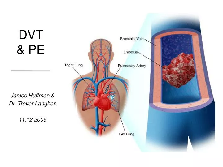
DVT & PE James Huffman & Dr. Trevor Langhan 11.12.2009
DVT • Diagnostic Algorithm • Determining Pre-test Probability • Useful Diagnostics • Treatment • Disposition • Special Circumstances • PE • Similar topics, PLUS: • Controversies
Virchow’s TriadWhite, RH: The epidemiology of venous thromboembolism. Circulation 107(23 Suppl 1):I4, 2003. • Injury to the vascular endothelium • Alterations in blood flow • Hypercoagulability Anything else associated with imbalanced clot formation? Age
Case 1 • 55♀: Referred to ED for pain, redness and swelling of the right calf • WIC today: Sent to ED with note:
Diagnostic Approach: Pre-test ProbabilityScarvelis, D., and P. Wells. 2006. Diagnosis and Treatment of DVT. CMAJ: 175(9); 1087
DVT: History & PE are Risk Assessment • Goals of H&P? • Determine pre-test probability • Look for other causes
History & Physical are Risk AssessmentAnand, SS, Wells, PS, et al. 1998. Does this Patient have deep vein thrombosis? JAMA:279(14)
DVT: H&P Bottom Line • Neither is sensitive or specific • i.e. you can’t rule-in or rule-out a DVT • Use them to decide pre-test probability
Pretest Probability • This algorithm re-presented in JAMA rational clinical examination series Anand SS, Wells PS, Hunt D, Brill-Edwards P, Cook D, Ginsberg JS. Does this patient have deep vein thrombosis? JAMA. 1998 Dec 2;280(21):1828-9. • What’s missing? The Dimer!
Assay Sensitivity Specificity Whole blood 80 - 85% 70 - 90% agglutination (SimpliRED) Latex 90 - 95% 40 - 90% agglutination R apid ELISA 95 - 100% 30 - 60% D-Dimer TestingKline, et al. Ann Emerg Med (2003); 42(2): 266-275. • Degradation product of fibrin • Non-specific • PPV bad • +ve: surgery, trauma, hemorrhage, CA, pregnancy, sepsis, >80 yrs old • Sensitivity variable • Need Pre-test probability to r/o DVT CLS uses
D-Dimer TestingWells, P., et al. 2003. NEJM: 349(13); pp1227-35 • RCT (N=1096) • D-Dimer vs no D-Dimer • DVT unlikely (Wells < 2) • # of U/S per pt decreased in D-dimer group (0.78 vs 1.34)
D-Dimer TestingWells, P., et al. 2003. NEJM: 349(13); pp1227-35 • “Modified” Wells Criteria • Used SimpliRED and IL-Test assays (less sens than ours) • Conclusion: • Wells <2 and negative D-Dimer can safely r/o DVT
Case 1 continued • Pretest probability? • Active cancer (1) • Localized tenderness (1) • Calf swelling (1) • Edema (1) • Other Diagnosis? Compression by pelvic nodes? (Doesn’t matter – score would still be “not low risk”) So she gets either 4 or 2 points = DVT likely
Level 2 – D-dimer • Her d-dimer was positive at 1.23
UltrasoundAmerican Journal of Respiratory Critical Care Medicine. 1999: 160; 1043-66 Bottom line: U/S is the test of choice for DVT
Anatomy Emerg Med Clin N Am. 26 (2008) • Depth: • Deep • Superficial* • Proximity: • Popliteal v. or higher • Distal *Superficial femoral vein is a member of the deep group
Ultrasound Fields, JM, & Goyal, M. Venothromboembolism. Emerg Med Clin of N Am. 2008; 26: 649-83
Bedside U/S? Jolly BT, et al. Acad Emerg Med 1997;4(2):129–32. Frazee BW, et al. J Emerg Med 2001;20(2):107–12.
Blaivas M, et al. Acad Emerg Med. 2000;7(2):120–6. • Median exam time of 3m 28s • 98% correlation with vascular lab-performed studies • Theodoro D, et al. Am J Emerg Med. 2004;22(3):197–200. • 125m reduction in time to pt disposition with EP-performed US • Kappa = 0.9, 99% agreement (154/156 cases) • Jang T, et al. Acad Emerg Med. 2004;11(3):319–22. • 8 emerg residents (4 PGY-1, 2 PGY-2, 2 PGY-3) • 1h focused training (didactic and practice on 2 healthy volunteers) • SN = 100%, SP = 91.8%, avg scan time = 11.7min (self-reported) • 4 false-positives (chronic DVT), 0 false-negatives
Ultrasound: Limitations • Obese, ++edema, immobilsation devices (x-fix) • Doesn’t see isolated thrombi in iliac or superficial femoral veins within abductor canal MRI better • Pelvic masses may cause noncompressibility in absence of thrombus false +’ve • Most importantly: U/S doesn’t return to normal after acute DVT • Therefore use impedance plethysmography for recurrent DVT • U/S - 60-70% of studies return to normal at one year • IP – 90% return to normal within a year
CT-VenographyGoodman LR, Stein PD, Matta F, et al. AJR Am J Roentgenol 2007;189(5): 1071–6
DVT: Bottom Line Thus Far? • Hx/PE help decide pretest probability (Wells) • Add a sensitive test (D-Dimer) • Almost all cases, do a sensitive confirmatory test (U/S)
Case 1 Continued • Okay back to it… • U/S shows popliteal vein DVT • Management Doctor?
Medical ManagementRosen’s Emergency Medicine 7th EditionScarvelis, D., and P. Wells. 2006. Diagnosis and Treatment of DVT. CMAJ: 175(9); 1087
Treatment: Bottom Line • IV UFH, LMWH, Fondaparinux are all acceptable
Case 1 Conclusion • Pt started on Enoxaparin and Warfarin • Arranged to see her oncologist and a hematologist as out-patient 2 days later • In General, discharge home is safe. • Admission may be required if: • Renal failure, high bleeding risk • Extensive DVT (painful blue leg) • Necessity for parenteral narcotics • Inability to have injections at home
Superficial ThrombophlebitisRosen’s Emergency Medicine 7th Edition • Uncommonly evolves into a thromboemboic event • BUT, ~8% of patients have synchronous DVT
Isolated Calf or Saphenous V. ThrombosisCanadian Medical Association Journal. 2003; 168(2) • Rarely cause significant PE • 25% of calf DVT extend to involve proximal veins • Vast majority will extend within 7d • DVT complications in 10-38% (untreated) Clinically this means you can Rx ASA (325mg/d) and arrange for re-U/S in 7d or just start on full DVT anticoagulation
Case 2 • 61♀ presents to ED complaining of mild pleuritic chest pain • Total knee arthroplasty 5/12 ago. Healthy otherwise
Risk AssessmentEmergency Medicine Reports. 2004;25(11) • History and Physical do not confirm the diagnosis, they merely raise the suspicion of the diagnosis, triggering further investigation • Hx: • Have to consider PE: dyspnea, tachypnea Pleuritic CP, syncope, hypotension & hemoptysis • Non-specific • PE: • Tachypnea and tachycardia are most common
Pretest Probability Emergency Medicine Reports. 2004;25(11) • All decision rules start w/ score • Wells and Geneva validated • Wells NPV: 99.5% • Others more cumbersome • Geneva (Wicki): adds ABG, CXR • PISA-PED: Adds ECG
Bottom Line: Use history & physical exam risk stratify patients Wells ≤ 4 PE Unlikely Wells > 4 PE Likely
H&P are Risk AssessmentWells, PS. J Thromb Haemost. 2007; 5(Suppl 1):41-50 Wells ≤ 4 Unlikely Wells > 4 Likely
Risk AssessmentEmergency Medicine Reports. 2004;25(11) • CXR: • Often AbN (Pleural effusion, atelectasis, elevated hemidiaphragm) • N CXR with dyspnea & hypoxemia = PE • Know Hampton’s and Westermark for exams • EKG: • Non-specific ST, Twave changes, Tachy • Signs of R heart strain (Anterior/Inferior T-wave inversions) • Know SIQIIITIII for exams • Simultaneous TWI in V1 and III are highly specific • ABG: • Hypoxemia common, but not always present • AAD02 >20 suggests PE (PIOPED) • 25-35% of pts with PE have normal blood gasses, pulse ox, and A-A gradient
Case 3 Continued • HR: 104 • Nil else • She gets 1.5 points • Now what? • Do you even start to work her up for PE?
PE Rule-Out Criteria (PERC Rule)Kline, JA. et al. J Thromb Haemost. 2004; 2:1247-55 • Based on the premise that overuse of D-dimer to screen for PE can have negative consequences • Derivation phase: • 3148 patients evaluated for PE in 10 US EDs • Data collected on 21 variables • Logistic regression and inter-observer agreement used to narrow to rule of 8.
PE Rule-Out Criteria (PERC Rule)Kline, JA. et al. J Thromb Haemost. 2004; 2:1247-55 • Age <50 • Pulse rate <100 beats/min • Oxygen saturation >94% • No hemoptysis • No unilateral leg swelling • No recent major surgery or trauma • No prior pulmonary embolism or deep venous thrombosis • No hormone use