Download

1 / 31
390 likes | 1.09k Views
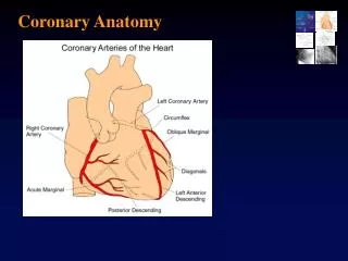
Coronary Anatomy, Variants and Lesion Characteristics. Ms. Leonardo Roever. Coronary Angiography. Pepine et al. Diagnostic and Therapeutic Cardiac Catheterization , 3 rd Ed. Routine Angiographic Views. Left Coronary Shallow RAO, AP Caudal LAO Caudal LAO Cranial RAO Cranial

E N D
Coronary Anatomy, Variants and Lesion Characteristics Ms. Leonardo Roever
Coronary Angiography Pepine et al. Diagnostic and Therapeutic Cardiac Catheterization, 3rd Ed.
Routine Angiographic Views Left Coronary • Shallow RAO, AP Caudal • LAO Caudal • LAO Cranial • RAO Cranial Right Coronary • LAO Cranial • AP Cranial, Shallow RAO • LAO Caudal
Optimal Coronary Angiographic Views: Left Main Segment Routine Adjunctive Ostial LAO Caudal AP Caudal Body RAO Caudal AP Caudal RAO Cranial Distal LAO Caudal LAO Cranial RAO Caudal
Optimal Coronary Angiographic Views: Left Anterior Descending Segment Routine Adjunctive Ostial LAO Caudal AP/RAO Caudal Body RAO Caudal AP Cranial LAO&RAO Cranial Distal RAO LAO Cranial RAO Caudal
Coronary Angiography Pepine et al. Diagnostic and Therapeutic Cardiac Catheterization, 3rd Ed.
Optimal Coronary Angiographic Views: Circumflex Segment Routine Adjunctive Ostial LAO Caudal AP Caudal Body Shallow RAO AP Caudal LAO LAO cranial Distal Shallow LAO LAO Cranial Shallow RAO AP caudal
Coronary Angiography Pepine et al. Diagnostic and Therapeutic Cardiac Catheterization, 3rd Ed.
Optimal Coronary Angiographic Views: Right Coronary Artery Segment Routine Adjunctive Ostial/Prox LAO Cranial Lateral(90-110) AP Caudal Mid Vessel Shallow RAO Lateral LAO Caudal Distal Bifurcation LAO Cranial AP Cranial PDA Shallow RAO AP Cranial PLB AP caudal Lateral Cranial
Coronary Angiography Pepine et al. Diagnostic and Therapeutic Cardiac Catheterization, 3rd Ed.
Coronary Angiography Pepine et al. Diagnostic and Therapeutic Cardiac Catheterization, 3rd Ed.
Coronary Angiography Lesion Classification: Coronary Angiographic Outcomes Predictors Based on AHA/ACC Grading System Type A Discrete Concentric Readily accessible Smooth Contour Little or no calcification Non ostial No major side branch involved Absence of thrombus
Coronary Angiography Lesion Classification: Coronary Angiographic Outcomes Predictors Based on AHA/ACC Grading System Type B Tubular Eccentric Moderate tortuosity Moderately angulated (45 – 900) Irregular contour Moderate – heavy calcification Total occlusion (< 3 months) Ostial Bifurcation lesion Thrombus present Note: B1 = characteristic only; B2 = 2 or more characteristics
Coronary Angiography Lesion Classification: Coronary Angiographic Outcomes Predictors Based on AHA/ACC Grading System Type C Diffuse Excessive Tortuosity Extremely angulated Total occlusion (> 3 months) Inability to protect major side branch Degenerated vein graft
Coronary Angiography Distal Blood Flow/Collateral Classification Based on TIMI Trial TIMI Grade Contrast Flow 0 (No perfusion) Antegrade flow to lesion; no flow beyond occlusion 1 (Penetration with Contrast passes beyond minimal perfusion) lesion but does not opacify distal vessel during cine run 2 (Partial perfusion) Contrast passes obstruction and fills distal vessel.However, rate of filling and/or washout slower than vessel segments outside lesion 3 (Complete perfusion) Contrast passes freely into distal at same visual rate as unaffected adjacent vessels
Coronary Angiography Distal Blood Flow/Collateral Classification Based on TIMI Trial Collateral Supply Contrast Flow 1 Absent 2 Minimal 3 Well developed Adapted from TIMI
Coronary Angiography NHLBI Classification System for Coronary Dissection DissectionA Small radiolucent area within the lumen of the vessel B Linear, nonpersisting extravasation of contrast C Extraluminal, persisting extravasation of contrast D Spiral-shaped filling defect E Persistent lumen defect with delayed anterograde flow F Filling defect accompanied by total coronary occlusion Length (in mm) Measure end to end for type B through F dissections Staining Persistence of contrast within the dissection after washout of contrast from the remaining portion of the vessel
Coronary Angiography Coronary Anomalies • Common: Insignificant • Conus Separate Ostium from RCA 50% • Left Circ from RCA 0.3% • Uncommon: Clinically Significant • LM from Proximal RCA • Courses between Great Vessels • Associated with sudden Death
Coronary Angiography Left Main Arising from RCA
Coronary Angiography Left Main from RCA Compression of LM Angulationat originof LM
Coronary Angiography Pepine et al. Diagnostic and Therapeutic Cardiac Catheterization, 3rd Ed.
Coronary Angiography LM-Right Sinus of ValsalvaSeptal Course Adapted from Seroth et al. Am J Cardiol 1990;65:891-898
Coronary Angiography LM-Right Sinus of Valsalva Retroaortic Course Adapted from Seroth et al. Am J Cardiol 1990;65:891-898
Coronary Angiography LM-Right Sinus of ValsalvaAnterior Free Wall Adapted from Seroth et al. Am J Cardiol 1990;65:891-898
Coronary Angiography LM-Right Sinus of Valsalva Interarterial Course Adapted from Seroth et al. Am J Cardiol 1990;65:891-898