Download
1 / 37
460 likes | 1.43k Views
MEDS 531 spring 2008 J.G. Seegmiller. Neck Viscera & clinical correlations of neck anatomy. Location. Viscera are located deep to the investing layer of cervical fascia & muscular triangle. NECK VISCERA FROM A CORONAL SECTION. POSTERIOR. Pretracheal fascia surrounds the neck viscera.

E N D
MEDS 531 spring 2008 J.G. Seegmiller Neck Viscera & clinical correlations of neck anatomy
Viscera are located deep to the investing layer of cervical fascia & muscular triangle
NECK VISCERA FROM A CORONAL SECTION POSTERIOR Pretracheal fascia surrounds the neck viscera ANTERIOR
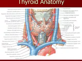
Cervical Viscera Layers • Respiratory Layer • Larynx • Trachea • Alimentary (Digestive) Layer • Pharynx • Esophagus • Endocrine layer • Thyroid • Parathyroids
Respiratory Layer • Larynx • Located between (C4-C6) • Easily palpated • Complex organ of voice production
Respiratory Layer • Trachea • Extending from larynx (C6 ) to right and left main bronchi • Tracheal cartilages 2-4 usually covered by isthmus • Inferior thyroid veins
Digestive Layer • Pharynx • Posterior to nasal and oral cavities • Ends at pharyngoesophageal junction
Digestive Layer • Cervical Esophagus • Upper third • Striated (voluntary muscle) • Between the trachea and cervical vertebrae Note relationship of esophagus & trachea to recurrent laryngeal nerve and paratracheal lymph nodes
Endocrine Layer • Thyroid (C5-C7) • Median isthmus (2-4 tracheal rings) • Lateral lobes • Superior and Inferior poles
Thyroid Variations No isthmus + pyramidal lobe Pyramidal lobe
Endocrine Layer • Thyroid Blood supply • Superior thyroid artery • Inferior thyroid artery • Sometimes thyroid ima artery Anterior view Posterior view
Endocrine Layer Venous drainage of the thyroid Thyroid veins Superior Middle Inferior To internal jugular veins To brachiocephalic veins
Endocrine Layer • Lymphatic drainage • Paratracheal nodes • Deep cervical nodes
Functions of the Thyroid Gland • Secretes thyroid hormones • Thyroxine & triiodothyronine • Maintains metabolic rate • Secretes calcitonin • Reduces blood calcium levels Follicular cells Thyroglobulin colloid Parafollicular (“C”) cells
Thyroid Gland Development • Originates in the back of the tongue • Migrates to the anterior neck Thyroglossal duct = A diverticulum of the embryonic pharynx (foregut)
Indicates the site of the orifice of the embryonic thyroglossal duct
Thyroid Pathologies • Goiter • Enlargement of the thyroid gland • Possible causes are numerous • Hereditary factors may cause goiters. Risk factors for the development of a goiter include female, age over 40 years, inadequate dietary intake of iodine, living in an endemic area, and a family history of goiter.
Thyroid Pathologies • Thyroglossal duct cysts • Remnants of epithelium remain
Thyroid Pathologies • Thyroglossal duct sinus
Thyroid Pathologies • Thyroid tumors • Benign (adenomas that secrete thyroid hormone) • Malignant (rare) Thyroid papillary carcinoma
Thyroid Pathologies • Ectopic thyroid tissue • Extra thyroid tissue may present anywhere along the path of the thyroglossal duct Lingual thyroid
Parathyroid Glands • Secretes parathyroid hormone • increases blood calcium levels • Stimulates osteoclasts to break down bone and release calcium • Increases gastrointestinal calcium absorption • Antagonist to calcitonin • Usually four
Parathyroid Development • Superior parathyroid • Derived from 4th pharyngeal pouch • Inferior parathyroid • Derived from the 3rd pharyngeal pouch
the operation or hindering the surgeon in any way. Intra-operative recurrent laryngeal nerve monitoring for the thyroid or parathyroid surgery
Retropharyngeal nodes Paratracheal nodes
Scheme for naming deep cervical node groups Jugulodigastric node Superior group Inferior group Supraclavicular nodes Jugulo-omohyoid node
Cancers originating from thoracic or abdominal organs can involve supraclavicular nodes
Clinical “zones” or “levels” of cervical nodes I – Submental and submandibular nodes. II – Upper deep cervical nodes. III – Middle deep cervical nodes. IV – Lower deep cervical nodes. V – Nodes in the posterior triangle. VI – Nodes in the muscular triangle.
Examples of regions/organs whose pathologies may present as enlarged nodes in these “levels”. Level II nodes Level III nodes Level IV nodes