Download
1 / 96
960 likes | 980 Views
Learn about Arboviruses: viruses transmitted by insect vectors, causing human diseases like yellow fever and Chikungunya. Discover their transmission modes, clinical manifestations, prevention methods, and laboratory diagnosis techniques.

E N D
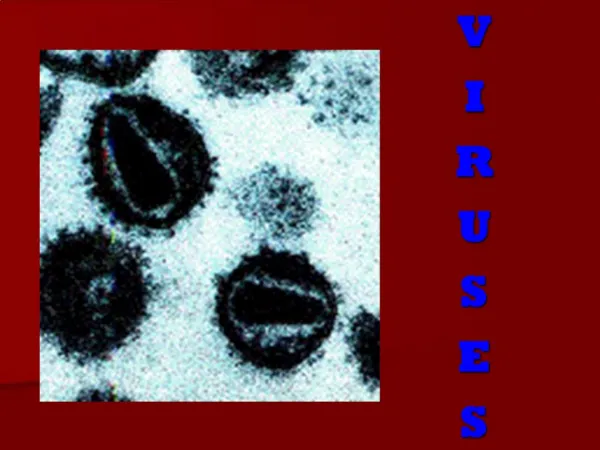
A R B O V I R U S E S ( Arthropod borne Viruses )
General Properties • Arbo viruses (Arthropod borne viruses) • Viruses of vertebrates biologically transmitted by haematophagus insect vectors. • They multiply in blood sucking insects and are transmitted to the vertebrate hosts. • Prevalent through out the world but are numerous in tropical than temperate climate. • About 500 viruses are there but in India 40 viruses have been detected. Many of them cause human disease.
Unaccepted taxonomically but useful biological concept. Named according to : Disease- yellow fever virus Place of isolation- Kyasanurforest disease (KFD) Local name of disease- Chikungunya Worldwide , Tropics India: > 40 Arbo viruses. Only > 10 cause disease.
“Hemotophagus” insect vectors . Vectors: Mosquitoes. Ticks. Phlebotomus Culicoide Less common Cimicidae
Placed in Toga viridae Flavi viridae. Bunya viridae Reo viridae Arena viridae. Filo viridae
Classification and structure A. Toga Viridae-alphavirus • Structure: Icosahedral, enveloped, 40-70 nm diameter, RNA
(D) Reo viridae • Structure: Helical, enveloped, RNA (ds RNA) • Members: • Colorado tick fever • African horse sickness • Palam, Kasba and Vellore viruses.
E)ARENA VIRIDAE Junin Machupo Lassa fever Lymphocytic choriomeningitis
F)FILOVIRIDAE Marburg Ebola
Mode of transmission: Insect Bite. Ingestion. Inhalation. Contact.
Pathogenicity • 3 main clinical entities • Fever with or without rashes • Hemorrhagic fevers • Encephalitis
Pathogenesis : Enters ‘ thru ‘ bite of insect Lymphatics Multiply RES (LN, liver, spleen , Endothelial cells of BV) Blood stream (Viremia) Target organs. CNSCapillary endotheliumLiver Multiplication Hemorrhagic Yellow fever Fever
Necrosis of Neurons, Perivascular cuffing of CBV, Sorrounded by microglia. Forms glial knots. ( Encephalitides )
Laboratory diagnosis : 1. Virus isolation: Patient Insects Réservoir animal Specimens : Blood, CSF. (During first 1- 3 days of illness)
Identification methods in Laboratory: 1. Suckling mice: Encephalitis 2. Chick embryo: CAM , Yolk sac. 3. Tissue cultures Primary: Chick embryo fibroblasts. Continuous cell lines: Vero / HeLa. Insect tissue.
4. Haemagglutination : Goose AND chick RBCs. 5. Serology : ELISA, CFT, HAI, Neutralization Test. 6. PCR technique : More sensitive. 7. Plaque reduction Neutralization Test (PRNT): More specific. “Antigenic” cross reactions among different groups.
Epidemiology : Zoonoses. Maintained in animals Except Dengue, O’ nyong nyong Asymptomatic in animals. Biting a viremic vertebrate . VectorsReservoir animal Human infection
Prevention : 1. Vector control. 2. Eradication of known reservoir. 3. Immunization : Vaccine Hyper immune Sérum
Togavirus : Enveloped, spherical, single stranded RNA viruses. Replicates in host cell cytoplasm. Alpha viruses All are mosquito borne HAI : Not specific Neutralisation test : More specific
Encephalitis Viruses :( America ) Eastern equine encephalitis Western equine encephalitis Venezuelan équine encephalitis Culex & Anophèles mosquitos. Wild birds are reservoirs.
Clinical manifestations: Sudden onset of fever Headache, Neck stiffness. Nausea, vomiting Disorientation Stupor or coma Convulsions in acute cases. Rigidity, weakness of limbs. Absent / irregular tendon reflexes.
CSF : Pleocytosis Protein, sugar levels are Normal. Identification of viral genome by PCR.
Febrile illness : With or without rash & arthralgia. CHIKUNGUNYA : 1952 Chikungunde = which folds up 1963 in India: As epidemics Calcutta, Madras Irregular outbreaks till 1973 in Maharashtra. Outbreak in 2006
Modes of transmission: 1.Transmitted exclusively by Aedes aegyptii. 2. Incubation period : 3 -12 days. 3 .Out breaks are common during the rainy season because of increased breading places for mosquito.
Clinical manifestations: Sudden onset of fever. Crippling Joint pains. Lymphadenopathy. Conjunctivitis. Maculopapular rash. Hemorrhagic manifestations rare. Biphasic fever typically
Diagnosis: 1. Clinical history. 2. Antigen detection (by using Monovalent antisera) 3. An IgM capture ELISA more useful. 4.A rapid, micro-scale focus reduction neutralization test for rapid diagnosis. 5. RT- PCR assay developed by Malaysians.
No vaccine. No specific therapy.
O nyong, nyong: Uganda. (Africa) Anopheles mosquito Resembles Chikungunya
Semliki Forest virus :Uganda, Aedes mosquito. Sindbis Virus : In India since 1952. Ross river virus : Epidemic polyarthritis in Australia.
Flaviviruses: Mosquito borne and tick borne Encephalitis viruses : Reservoirs :Wild birds. Vector : Culex St. Louis encephalitis virus in USA West Nile virus: India (Karnataka) Dengue like disease Murray Valley encephalitis virus in Australia.
Japanese ‘B’ encephalitis: Japan 1871. Isolated in 1935. Summer and autumn. Vector:Culex tritaeniorhychus Human infection is Dead-end event. Reservoir host : Herons. Amplifier Host : Pig.
Clinical Features: Abrupt onset of fever, Headache, Vomiting. Signs of encephalitis: Nuchal rigidity. Convulsions, confusion. Dysarthria, Tremors. Altered sensorium. Coma.
Pathogenesis : I phase : Viral multiplication in neuronal tissue. Seen in blood 3 days before CNS involvement. II phase: Major illness Viral multiplication in CNS Injury Destruction leads to
Lesions in basal structures & Cerebral cortex. Small hemorrhages. Perivascular cuffing. Meningeal infiltration Nerve damage. Patches of encephalomalacia. Spongiform appearance.
Lab diagnosis : Best infirst 1 - 3 days of illness Peripheral Smear: Neutrophilia CSF : Pleocytosis ( Lymphocytes ) Viral isolation.
Epidemiology : Usually Asymptomatic. 500 – 1000 unapparent infections /every case. General Mortality is 50% . In the old age 80%. Residual neurological damage : 50%
India : 1955 from Vellore. Children. Epidemics October and November. South India (AP ,TN ) only up to 1973. 1976 Dibrugarh. Gorakhpur Kolar
Prevention: 1. Mosquito control. 2. Vigilance on Piggeries. 3. Hyper immune serum: 1 - 2 days after infection. 4. Vaccine
Vaccines : 1.Formalin inactivated mouse Brain Vaccine. Nakayama strain Two doses : 2 weeks interval Booster : 6 – 12 months. Immunity is short lived. 2.Primary Baby Hamster kidney cell Live attenuated vaccine. JE strain SA 14-14-2 2 doses . 1 year apart 3. Vaccination of pigs
Hemorrhagic fevers : Dengue (Types 1 – 4) Aedes aegyptii Yellow fever Kyasanur Forest disease Ixodid ticks Omsk hemorrhagic Fever
Hemorrhagic fever with systemic involvement : YELLOW FEVER : Yellow Jack in Africa Epidemics : Central America. Caribbean New York Vector : Aedes aegyptii Extrinsic IP : 12 days Incubation period: 3 - 6 days.
Clinical Features:Acute fever, Chills, Backache. Nausea, Generalized malaise, Vomiting Slow pulse rate, temperature. Jaundice, Albuminuria, Hemorrhagic manifestations on mucosa.
Histologically : Liver : Fatty degeneration Mid zonal necrosis. Intracytoplasmic inclusions: Councilman bodies. Kidney :Focal Degeneration of tubular epithelium. Spleen & LN : Focal Degeneration. CNS : Intranuclear acidophilic inclusion bodies in Nerve, Glial cells-- “Torres bodies.
Epidemiology : Unapparent infections common. Not present in India . All ages between 15 – 45. Mild in infants than old age. Monkey is reservoir. Wood cutters, Nut pickers, Road builders : Usual victims
Urban cycle and sylvatic cycle • Urban cycle • Humans – both definitive host & reservoir • Aedes aegypti – vector • Sylvatic cycle • Wild monkeys – reservoirs • Haemagogus spegazzinii & Aedes africanus
Control : 1. Mosquito control. 2. Vaccines : a. French neurotropic vaccine Administered : By scarification. Complication: Encephalitis.
B. 17D live attenuated vaccine. Chick embryo ( Yolk sac ). Asibi strain Mouse embryo. Route of Administration : Sub cutaneous 0.5 ml. Validity : 10 yrs. Administered from 9 months of age.