Ultrasonic Imaging Parameters
Ultrasonic Imaging Parameters. Student: Mei-Ru Yang Wei-Ning Lee Advisor: Pai-Chi Li. Blood velocity. S. K. Alam and K. J. Parker,”The butterfly search technique for estimation of blood velocity,” Ultras. in Med. Biol.vol. 21, pp. 657-670, 1995.

Ultrasonic Imaging Parameters
E N D
Presentation Transcript
Ultrasonic Imaging Parameters Student: Mei-Ru Yang Wei-Ning Lee Advisor: Pai-Chi Li
Blood velocity S. K. Alam and K. J. Parker,”The butterfly search technique for estimation of blood velocity,” Ultras. in Med. Biol.vol. 21, pp. 657-670, 1995.
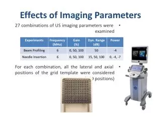
Evaluation of estimation techniques • Computational complexity • SNR • Blood backscatter is weak • The estimation technique must perform well in noisy situations. • The number of successive scan lines • This relates to the color flow imaging rate
The butterfly search technique • Can overcome the tradeoff criterion between image resolution and velocity resolution. • Combines some of the best features of time domain and Doppler methods. • Is reliable in hardware without exensive correlation calculations.
The envelope of echoes from a single reflector movement Movement of envelope on the time frame with the movement of the scatter t Fast time (range) Slow time (index) RF pulse envelope n
movement t Fast time (range) Slow time (index) RF pulse envelope n Butterfly lines signifying different velocities RF or envelope search -- a time domain technique • If the trajectory matches the scatter movement, all the data samples would have the same value and their variance will be zero. • Noise • variance is minimum 2d/c vo
Year US5086775 Paper 4 Paper 1 Paper 2 US5632277 Paper 3 Company (school) Ruhr University Rochester University Siemens Medical Systems California University
Nonlinear ultrasonic imaging B. H. H. and R. Y. C., “Ultrasound Imaging With Higher-Order Nonlinearities” , US patent 6063033, May 16, 2000.
Introduction • Fundamental and harmonic signal components • Direct echoes of the transmitted pulse • Generated in a nonlinear medium ex. tissue • Second harmonic • Worse SNR compared with fundamental • Improve image quality (Clutter rejection ) • Higher harmonic • SNR ???
frequency -fo fo B B frequency -2fo 2fo 2B -3fo 3B -fo 3B fo 3B 3fo 3B frequency Separate the second harmonic from the fundamental frequency • Bandpass filter • A transmitted signal centered at fo • The receive filter is centered at 2fo • Challenge : bandwidth requirement passband : (fo-B/2) to (2fo+B)
Method to extract the higher order nonlinear components Modelthe nonlinear echo signal polynomial expansion of some basis waveform Solve the coefficient of this model (least squares inversion)
Transmit signal pi(t) = bi po(t) i = 1….I bi : the same waveform with different complex amplitude received echo signals complex (B) excitation harmonic components
System Nonlinearities • Odd order system nonlinearities of circuit • Voltage symmetric pulser and amplifier • To cancel the system nonlinearities • by subtracting the linear echo
Year US5577505 US5632277 Paper 5 US6063033 US6344023 US6364836 Company (school) Siemens Medical Systems GE Hewlett Packard Matsushita Electric Industrial
Elastic modulus • Elastic constant • Young’s modulus, shear modulus, bulk modulus • Young’s modulus • For soft tissue E=3μ
Device Circuit Method Apparatus System Process Total Elastic modulus 2 0 16 6 0 3 15 Total amount 2 0 16 6 0 3 標的分析統計表
Year 1990 Paper 10 PN/ 4,947,851 1994 Paper 9 1995 Paper 7 1996 Kernel paper Paper 6 PN/ 5,495,777 PN/ 5,524,636 1999 Paper 8 Enterprise US Japan USSR Fujitsu ACAL 引證族譜圖
Reconstruct elastic modulus [1] • Elasticity imaging • Measurement of tissue displacement using speckle tracking algorithm • Measurement of strain tensor component • Spatial distribution of the elastic modulus using strain images
Cont’d Constitutive equation Static equilibrium equation
Cont’d Relation of two RF images envelop intensity f2(x, y)=(1-k)‧f1(x-ux, y-uy) Static equilibrium eq: Taylor expansion (f2-f1)+uxfx+uyfu+kf1=0 The least squares estimation Minimize Finite element method Estimate 2-D displacement(ux, uy) distribution Spatial derivative Strain tensor Shear modulus
Implement • Make a gel-based phantom • Select ROI • Spatio-temporal derivative method
References • Paper 1:S. K. Alam and K. J. Parker,”The butterfly search technique for estimation of blood velocity,” Ultras. in Med. Biol.vol. 21, pp. 657-670, 1995. • Paper 2: S. K. Alam and K. J. Parker,” Reduction of comptational complexity in the butterfly search technique,” IEEE Bio. Eng., Vol. 43, no. 7, 1996. • Paper 3:M. Vogt, “Application of high frequency resolution blood flow measurement,” IEEE Ultra. Symp. Pp.12431246, 1997. • Paper 4: K. W Ferrara, “A new wideband spread target maximum likelihood estimator for blood velocity estimation. II. Evaluation of estimator with experimental data” IEEE UFFC,Vol 33, 1991. • Paper 5: B. H. H. and R. Y. C., “Higher-order nonlinear Ultrasound Imaging ” ,IEEE Ultra. Symp.,1999.
Cont’d • Paper 6: Yasuo Yamashita, Misuhiro Kubota,“Tissue elasticity reconstruction based on ultrasonic strain measurements,” IEEE ultrasonic symposium, 1996. • Paper 7: A. R. Skovoroda, S.Y. Emelianov, and M. O’Donnell, “Tissue elasticity reconstruction based on ultrasonic displacement and strain images,” IEEE Trans. Ultrason., Ferroelect, Freq. Contr., vol.42, pp. 747-765, July 1995. • Paper 8: A. R. Skovoroda, Mark A. Lubinski, S.Y. Emelianov, and M. O’Donnell, “Reconstructive elasticity imaging for large deformations,” IEEE Trans. Ultrason., Ferroelect, Freq. Contr., vol.46, No. 3, May 1999. • Paper 9: A. R. Skovoroda, S.Y. Emelianov, Mark A. Lubinski, A. P. Sarvazyan and M. O’Donnell, “Theoretical analysis and verification of ultrasound displacement and strain imaging,” IEEE Trans. Ultrason., Ferroelect, Freq. Contr., vol.41, pp. 302-313, May 1994. • Paper 10: J. Ophir, I. Cespedes, H. Ponnekanti, Y. Yazdi, and X. Li,” Elastography: a quantitative method for imaging the elasticity of biological tissues,” Ultrason. Imaging, vol. 13, pp.111-134, 1991