
Animal structure and function
E N D
Presentation Transcript
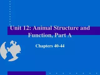
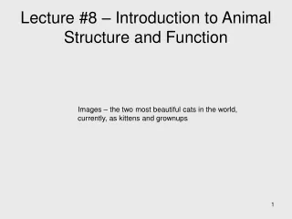
0 Forearm Wrist Finger 1 Palm Shaft Internal bone structure Finger 2 Finger 3 Shaft Barb Featherstructure Barbule Hook Form and Function • Functions result from specific structures Figure 20.1
Hierarchy of Structure 0 Cells -> Tissues -> Organs -> Organ system -> Organism A Cellular level Muscle cell B Tissue level Muscle tissue C Organ level Heart D Organ system level Circulatory system E Organism level Many organ systemsfunctioning together Figure 20.2A–E
Tissues • Groups of similar cells, same function • Types • Epithelial • Connective • Muscle • Nervous
Epithelial tissue • Functions: protection, secretion, and exchange 0 Free surface ofepithelium Basementmembrane(extracellularmatrix) Underlyingtissue Cellnuclei A Simple squamous epithelium(lining the air sacs of the lung) D Stratified squamous epithelium(lining the esophagus) Layers ofdead cells B Simple cuboidal epithelium(forming a tube in the kidney) Rapidly dividingepithelial cells Colorized SEM E Stratified squamous epithelium(human skin) C Simple columnar epithelium(lining the intestine) Figure 20.4A–E
0 Fatdroplets Cartilage-formingcells C Adipose tissue Matrix Cellnucleus D Cartilage (at the end of a bone) Collagenfibers Centralcanal B Fibrous connective tissue (forming a tendon) White bloodcells Cell Matrix Collagenfiber Bone-formingcells Red bloodcell Elasticfibers E Bone Plasma A Loose connective tissue (under the skin) F Blood Connective tissue Functions and types: ? Loose CT, adipose, blood, fibrous, bone, cartilage Figure 20.5A–F
0 Musclefiber Unit ofmusclecontraction Junction betweentwo cells Musclefiber Nucleus Nucleus Muscle fiber Nucleus B Cardiac muscle A Skeletal muscle C Smooth muscle Muscle tissue Functions and types: Skeletal, smooth and cardiac Figure 20.6A–C
Cell body Nucleus Cell extensions LM 330 Figure 20.7 Nervous tissue • Communication network • Neurons transmit nerve signals • Electrical and chemical 0
Lumen Small intestine(cut open) Lumen Epithelial tissue(columnar epithelium) Connective tissue Smooth muscletissue (2 layers) Connective tissue Epithelial tissue Organs 0 • Made of several tissues Figure 20.9
Organ systems • May have multiple functions • 11 systems in humans
Nasal cavity Mouth Larynx Esophagus Trachea Liver Bronchus Stomach Smallintestine Lung Largeintestine Anus B Respiratory system A Digestive system Digestive and respiratory systems Figure 20.10A, B
Bonemarrow Heart Thymus D Immune system Spleen E Lymphatic system Lymphnodes Bloodvessels Lymphvessels C Circulatory system Circulatory and lymphatic (immune) system • Moves food and oxygen • Protection Figure 20.10C–E
Pituitary gland F Excretory system Thyroid gland Thymus Kidney Adrenal gland Pancreas Ureter Testis(male) Urinarybladder Urethra Ovary(female) G Endocrine system More systems • Excretory • Endocrine and nervous systems • Control body functions Figure 20.10F–G
Hair Cartilage Skin Skeletalmuscles Nails Bones J Skeletal system I Integumentary system K Muscular system More Systems • Integumentary system • Skeletal and muscular systems Figure 20.10I–K
Male Female Prostategland Vasdeferens Oviduct Ovary Urethra Uterus Penis Vagina Testis L Reproductive systems Reproductive system Figure 20.10L
Seeing Inside • Computed tomography (CT) scans Figure 20.11A Figure 20.11B
MRM • Magnetic resonance microscopy (MRM) 3D! Figure 20.11C
MAX HEARINGWORDS SEEINGWORDS MIN GENERATINGWORDS SPEAKINGWORDS • PET • Positron-emission tomography (PET) Figure 20.11D
Interactions with External Environment • Open systems: • Exchange between animals and environment
0 Mouth Diffusion Twocelllayers Diffusion Gastrovascularcavity Interactions with External Environment • Small animals • Use surface area to meet their cells’ needs Figure 20.12A
External environment CO2 O2 Food Mouth Animal Respiratorysystem Digestivesystem Interstitialfluid Nutrients Circulatorysystem Bodycells Excretorysystem Intestine Anus Metabolic wasteproducts (urine) Unabsorbedmatter (feces) Interactions with External Environment • Larger animals • Specialized structures to increase surface area • Exchange in interstitial fluid Figure 20.12B
Larger animals Figure 20.12C
Externalenvironment Internalenvironment Homeostaticmechanisms Smallfluctuations Largefluctuations Homeostasis • Regulation of internal environment • Staying within safe levels • Maintain constant internal environment Figure 20.13A Figure 20.13B
Negative Feedback Figure 1.5
Positive Feedback • output enhances or exaggerates the original stimulus Figure 1.6
Thermoregulation • Ectotherms • Endotherms
LE 40-12 40 River otter (endotherm) 30 Body temperature (°C) 20 Largemouth bass (ectotherm) 10 0 10 20 40 30 Ambient (environmental) temperature (°C)
Thermoregulation • Heat loss/gain • Integumentary system • Evaporation • Behavior • Vasodilation/-constriction