Understanding Adhesive Capsulitis: Risk Factors, Diagnosis, Treatment
250 likes | 342 Views
Learn about adhesive capsulitis, including risk factors, evaluative tools for diagnosis, phases, and treatment ideas. Understand the anatomy, predisposing factors, pathophysiology, differential diagnosis, stages, evaluation, signs, symptoms, outcome measures, conservative interventions, and physical therapy strategies.

Understanding Adhesive Capsulitis: Risk Factors, Diagnosis, Treatment
E N D
Presentation Transcript
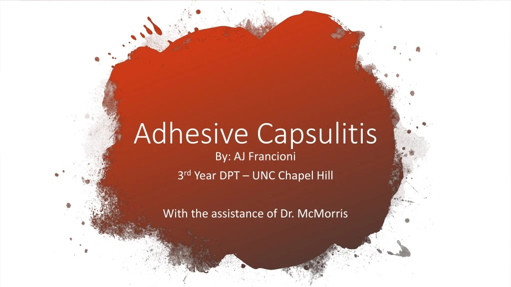
Adhesive Capsulitis By: AJ Francioni 3rd Year DPT – UNC Chapel Hill With the assistance of Dr. McMorris
Learning Objectives • Recognize at least 3 risk factors for Adhesive Capsulitis • Recognize at least 3 evaluative tools to diagnose Adhesive Capsulitis • Be able to identify the phases of Adhesive Capsulitis • Be able to identify at least 3 treatment ideas for Adhesive Capsulitis
Adhesive Capsulitis • GHJ capsular fibrosis with chronic inflammation • Primary • No specific, precipitating event • (Believed) chronic inflammation with fibroblastic proliferation • Secondary • After surgery or injury • Potential associated conditions • Diabetes, RC injury, CVA, cardiovascular disease, thyroid • Incidence = 5-percent of general population1 • As much as 20-percent for people with diabetes1
Anatomy to Consider2 • GHJ encased by capsule, which has 2 layers • External = dense, fibrous connective tissue • Inner = protein collagen • Synovial membrane • if fluid is underproduced ROM loss • Capsule is strongest superiorly restricts rotation • Ligaments thicken capsule anteriorly restricts external rotation • Capsular Pattern = ER > AB > IR • Common finding through imaging = coracohumeral ligament becomes stiffer
Postero-Inferior view of shoulder dissection to demonstrate the anterosuperior glenohumeral capsule.3
Predisposing Factors • Middle age (40-59 years) 4 • Female4 • Diabetes Mellitus1 • Thyroid disease1 • Trauma1 • Autoimmune disease1 • Cerebrovascular disease, CAD, MI1 • Sedentary lifestyle5 • Past h/o Adhesive Capsulitis • Prolonged immobilization2 • Patient Education • Modifiable Risk Factors • Level of Complexity • Adherence to HEP
Pathophysiology • Inflammation + Fibrosis = pain and stiffness • Accepted pathology • Contracture of GH capsule1 • Loss of synovial layer1 • Adhesions of axillary tissue to itself1 • Decreased capsular volume1 • Fascial restrictions, muscle tightness, and trigger points • Current Hypothesis = inflammation in joint capsule and synovial fluid allow for fibrosis and adhesions in synovial lining4
Glenohumeral capsule during the “frozen” phase of adhesive capsulitis6
Differential Diagnosis1,4 • Septic arthritis • Fracture • Rotator Cuff pathology • GH arthrosis • Shoulder impingement • Cervical radiculopathy • Chronic Regional Pain Syndrome • Shoulder Girdle tumors • Tendonitis/bursitis • Fibromyalgia • Disease of digestive system
Four Stages • Stage 1 = up to 3 months • Stage 2 = Painful “Freezing” Stage lasts from 3-9 months4 • Stage 3 = “Frozen” Stage lasts from 9-15 months4 • Stage 4 = “Thawing” Stage lasts from 15-24 months4
Stage 1 • Up to 3 months • Sharp pain at end of ROM • Can be mistaken for impingement d/t greater motion still available • Achy pain at rest • Sleep disturbance
Stage 2 – Freezing • Lasts 3 to 9 months1 • Inflammation/synovitis1 • Present with diffuse shoulder pain or stiffness4 • More active at night • Gradual loss of motion d/t pain
Stage 3 - Frozen • Lasts 9 to 15 months4 • Pain and ROM loss d/t adhesions and synovial proliferation1 • Capsular Pattern = ER > AB > IR Stage 4 – Thawing • Lasts 15-24 months4 • Recovery phase with gradual return of ROM • 20-50 percent of patients will have lasting symptoms past this phase1 • Pt education for adherence and follow-through
Evaluation and Examination • Objective • Clear cervical spine • Active and Passive ROM • Compare sides • Strength • “Shrug sign” with GH elevation • Special Tests:4 • Neer • Hawkins-Kennedy • Outcome Measures Signs and Symptoms • Gradual onset of pain progressively worse • Guarding or protect it by reducing movement • Difficulty with UE focused tasks • Sleep disturbance • Night pain
Outcome Measures7 • Self-reported measures asking about ADL’s and functional tasks • Disabilities of the Arm, Shoulder, and Hand (DASH) • American Shoulder and Elbow Surgeons shoulder scale (ASES) • Shoulder Pain and Disability Index (SPADI) • Reaching overhead • Sleeping on affected side • Washing hair • Carrying heavy object
Conservative Interventions • NSAIDS • Oral corticosteroids • Modalities to relieve inflammation • Physical therapy
Physical Therapy Painful “Freezing” Stage • PROM to maintain existing ROM and relieve muscle involvement6 • Postural positioning8 • Grade I/II mobs and long axis distraction1,4
Physical Therapy Frozen and Thawing • AAROM8 • Capsular stretching1 • Progressive resistance training in pain-free range4 • HEP • Stretching8 • Scapular and RC strengthening
Referrals and Discharge • Orthopedic specialist • No improvements of symptoms or functional mobility within 6 months9 • Educate patient on expectations • Co-morbidities can affect perceived outcomes and timeliness of progress • Long-term disability 10-20 percent4 • Persistent Symptoms 30-60 percent4 • Educate on importance of persistence with HEP • Consider the biopsychosocial model when treating
Procedural Interventions • GH intra-articular corticosteroid injection • Fast relief by reducing inflammation, but effects may only last 4-6 weeks 7 • Hydrodilation • Inject large volume of fluid to increase intracapsular volume and stretch capsule 1,4,6 • Manipulation under anesthesia • Capsule or scar tissue stretches or tears6 • Arthroscopic surgery • Risk d/t period of immobilization required6
The Interactive Part of Learning Objectives • List at least 3 risk factors for Adhesive Capsulitis • List at least 3 evaluative tools to diagnose Adhesive Capsulitis • Explain the phases of Adhesive Capsulitis • Provide at least 3 treatment ideas for Adhesive Capsulitis
References • Le HV, Lee SJ, Nazarian A, Rodriguez EK. Adhesive capsulitis of the shoulder: review of pathophysiology and current clinical treatments. Shoulder Elbow 2017;9(2):75-84. doi:10.1177/1758573216676786. • Carmichael SW, Hart DL. Anatomy of the shoulder joint. J. Orthop. Sports Phys. Ther. 1985;6(4):225-228. doi:10.2519/jospt.1985.6.4.225. • Duke Anatomy - Lab 16: Upper & Lower Limb Joints. Available at: https://web.duke.edu/anatomy/lab16/lab16.html. Accessed December 4, 2018. • St Angelo JM, Fabiano SE. Adhesive Capsulitis. In: StatPearls. Treasure Island (FL): StatPearls Publishing; 2018. • Rauoof MA, Lone NA, Bhat BA, Habib S. Etiological factors and clinical profile of adhesive capsulitis in patients seen at the rheumatology clinic of a tertiary care hospital in India. Saudi Med J 2004;25(3):359-362. • Frozen Shoulder - Adhesive Capsulitis - OrthoInfo - AAOS. Available at: https://orthoinfo.aaos.org/en/diseases--conditions/frozen-shoulder/. Accessed December 4, 2018. • Kelley MJ, Shaffer MA, Kuhn JE, et al. Shoulder pain and mobility deficits: adhesive capsulitis. J. Orthop. Sports Phys. Ther. 2013;43(5):A1-31. doi:10.2519/jospt.2013.0302. • Chan HBY, Pua PY, How CH. Physical therapy in the management of frozen shoulder. Singapore Med J 2017;58(12):685-689. doi:10.11622/smedj.2017107. • Neviaser AS, Neviaser RJ. Adhesive capsulitis of the shoulder. J Am AcadOrthopSurg 2011;19(9):536-542.