Download
1 / 17
170 likes | 347 Views
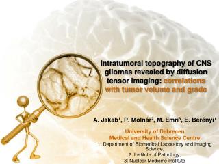
Intratumoral topography of CNS gliomas revealed by diffusion tensor imaging: correlations with tumor volume and grade. A. Jakab 1 , P. Molnár 2 , M. Emri 3 , E. Berényi 1 University of Debrecen Medical and Health Science Centre 1: Department of Biomedical Laboratory and Imaging Science,

E N D
Intratumoral topography of CNS gliomas revealed by diffusion tensor imaging: correlations with tumor volume and grade A. Jakab1, P. Molnár2, M. Emri3, E. Berényi1 University of Debrecen Medical and Health Science Centre 1: Department of Biomedical Laboratory and Imaging Science, 2: Institute of Pathology, 3: Nuclear Medicine Institute
…the mobility of water molecules may be considered to change with the progression of normal tissue to a state of disease. These changes in the mobility of water molecules are "fingerprinted" by changes in the molecular motion of the solids. Carlton F. Hazlewood Baylor College of Medicine from 1965 until December 31, 1997, professor of Molecular Biology and Biophysics. Hazlewood CF. Information forgotten or overlooked: fundamental flaws in the conventional view of the living cell. Cell Mol Biol (Noisy-le-grand). 2001 Jul;47(5):959-70. Review. A. Jakab, E. Berényi | DE OEC OLKD | Symposium Neuroradiologicum
5 Introduction Imaging diffusion • Diffusion in biological tissues isrestricted, anisotropic • Diffusion tensor imaging (DTI)depicts tissue anisotropy • Alteration in diffusion characteristics can be brought about by: • disorganisation of the ECM • changes in citoskeletal elements • necrosis • new vessel growth? • biological behaviour of higher grade? (e.g. more proliferations, different cellularity) A. Jakab, E. Berényi | DE OEC OLKD | Symposium Neuroradiologicum
6 Introduction Diffusion metrics for tumor grading • DTI for assessing physiological and pathological parameters (current literature): • Fractional anisotropy: tumor cellularity and proliferation rate • MRS and DTI: proliferation rate • FA: Low grade vs. Anaplastic glioma • ADC histograms: 1p/19q l.o.h. in oligodendrogliomas • Visualization of the „Oligo-like” and „astro-like” component [Khayal et al., 2009] • THEREFORE diffusion tensor measurements may beuseful in determining tumor grade: • Glioma grading by ADC and FA values[Lee et al., 2008] • Glioma grading by histogram analysis of DTI scalars[Jakab et al., 2010] A. Jakab, E. Berényi | DE OEC OLKD | Symposium Neuroradiologicum
7 Introduction Tumor growth • Disordered – ordered diffusion? intratumoral space revealed by diffusion tensor imaging (glyph colour: direction of diffusion) A. Jakab, E. Berényi | DE OEC OLKD | Symposium Neuroradiologicum
8 Objectives • Our hypotheses and aims: • Grade is reflected by diffusion characteristics • Tumor growth, tumor size, neovascularization, and volume of the necrotic core affects diffusion characteristics or ALTERS the results of measurements like fractional anisotropy or ADC • Our purpose was to measure diffusion values of tumors of different size by designating regions such as tumor center and tumor periphery A. Jakab, E. Berényi | DE OEC OLKD | Symposium Neuroradiologicum
9 Methods Patient population, imaging • Retrospective evaluation of patients with proved pathological diagnoses • First cohort: 25 gliomas, (9 high grade, 16 low grade) • Second cohort: only diffuse astrocytomas (13 WHO grade II) • Diffusion tensor imaging (DTI) protocol • GE Signa Excite Twingradient, 1.5T • SS EPI, MPG: 25 directions, b-value=1000 s/mm2TR / TE: 1000 / 98 ms; voxel size: 1 x 1 mmSlice thickness: 3.3 mm A. Jakab, E. Berényi | DE OEC OLKD | Symposium Neuroradiologicum
10 Methods Region analysis • 2 tumor regions: tumor periphery and tumor center • Periphery: high signal intensity abnormality on B0 images • Center: low signal intensity abnormality on FA images HIGH GRADE LOW GRADE A. Jakab, E. Berényi | DE OEC OLKD | Symposium Neuroradiologicum
11 Methods Topography index, variables Variables in analysis: Gross tumor volume (blue line) c= centerb= border cFA, bFAcADC, bADCcDWI, bDWIFA index: cFA / bFAiDWI index: cDWI / bDWIADC index: cADC / bADC cFA bFA A. Jakab, E. Berényi | DE OEC OLKD | Symposium Neuroradiologicum
12 Results Tumor volume and topography index ONLY THE cFA / bFA index SHOWED CORRELATION WITH TUMOR SIZE A. Jakab, E. Berényi | DE OEC OLKD | Symposium Neuroradiologicum
13 Results Tumor volume and topography index THE CORRELATION BETWEEN THE ANISOTROPY TOPOGRAPHY INDEX WAS NEARLY IDENTICAL IN LOW- AND HIGH-GRADE GLIOMAS A. Jakab, E. Berényi | DE OEC OLKD | Symposium Neuroradiologicum
14 Results Tumor volume and topography index Correlation between tumor volume and FA topography index A. Jakab, E. Berényi | DE OEC OLKD | Symposium Neuroradiologicum
15 Results Tumor volume and topography index Correlation between tumor volume and FA topography index: ONLY DIFFUSE ASTROCYTOMAS = higher correlation: Pearson: -0.75, p=0.003 A. Jakab, E. Berényi | DE OEC OLKD | Symposium Neuroradiologicum
16 Discussion • Structural and diffusion features of tumor core differ fromthose of the periphery. • The degree of anisotropy is correlated with tumor size after defining a center to periphery index. • There is a tendency towards lower FA in the tumor core compared to tumor periphery. • These observations require validation based on increased sample size in each categories. • Practical considerations: • 1. Tumor size as a coefficient in grading tumors with diffusion measurements • 2. More data may reveal such intratumoral characteristics which might permit predictions vis-á-vis drug delivery and distribution! A. Jakab, E. Berényi | DE OEC OLKD | Symposium Neuroradiologicum