Download
1 / 34
350 likes | 426 Views
Learn about the vital factors for spermatogenesis, process of sperm cell production, erection and ejaculation mechanisms, ovulation induction process, development of male gender, hormone production in testis, and androgen effects on development.

E N D
Most important points Fonyó: Orvosi Élettan, Medicina, Budapest, 1997, Fig. 33-4. • factors needed for spermatogenesis: FSH, testosterone (LH), low temperature, vitamin A • sperm cells are continuously produced from stem cells • spermatogonium (2n) – primary spermatocyte (4n) –secondary spermatocyte (2n) – spermatid (1n) • erection is parasympathetic, ejaculation is sympathetic process, NO has a crucial role (see Viagra) • women have the total set of primary oocytes (4n) after birth – risk for adverse environmental effects is higher • follicles mature from primary to quaternary (Graafian) in about 200 days (misleading textbook figures!) • secondary oocytes (2n) are formed during ovulation, reduction of chromosome number to 1n occurs during fertilization • ovulation is induced by an estrogen peak, followed by an LH peak supported also by progesterone (positive feedback) • corpus luteum kills itself by exerting negative feedback on LH production – in pregnancy hCG saves it • delivery is induced by the baby’s adrenal gland switching from the production of androgens to glucocorticoids – estrogen is produced in the placenta testicle epididymis 2/24
Development of male gender Fonyó: Orvosi Élettan, Medicina, Budapest, 1997, Fig. 33-4. • genetic sex is determined at the moment of conception by X and Y chromosomes • development of gonadal sex requires the presence of male or female gonads, depending on the presence of testis-determining-factor • intrauterine and post-natal development of phenotypic sex depends on hormones produced by the gonads • these sex hormones also influence the development of the CNS – psychological sex • the male gonad, the testis consists of curled tubules (seminiferous) • in the wall of the tubules Sertoli-cells (nurse-cells) are found, their function is the spermatogenesis; between the tubules Leydig cells synthesize the gonadal hormones testicle epididymis 3/24
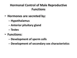
Hormone production in the testis Eckert: Animal Physiology, W.H.Freeman and Co., N.Y.,2000, Fig. 9-30. • the major steroid produced by the testis testosterone, it is transformed into dihydrotestosterone in the targets cells – it is the most effective androgen • the necessary cholesterol is provided by LDL particles and by de novo synthesis (50-50%) • testosterone is transported in blood by testosterone-binding globulin (50%) and albumin (50%) • effects of the hormones • prenatal life: gonadal and phenotypic sex • during puberty: male genitalia, secondary sexual marks • after puberty: spermatogenesis and preservation of sexual marks • steroid synthesis is facilitated by LH (ICSH) through cAMP • LH acts on functioning and synthesis of enzymes • androgens (and oestradiol) inhibit GnRH, LH and FSH; inhibin from Sertoli-cells FSH 4/24
Androgens and development I. Berne and Levy, Mosby Year Book Inc, 1993, Fig. 51-6 • after the 13 intrauterine week (second trimester) high LH and FSH level, Leydig-cells produce testosterone (maternal hCG also facilitates), no feedback regulation yet – hormonal concentration approaches adult levels • testosterone causes differentiation of the same group of cells into male genitalia that form female genitalia in girls • hormone level is low in the last trimester, but there is a few-month long postnatal peak in the first year – function unknown • around the age of 6-7 androgen production in the adrenal cortex increases adrenarche – hair growth on limbs and around genitalia, acceleration of growth • after the age of 12 hormone production in the testis starts to increase reaching its maximum in a few years 5/24
Androgens and development II. • androgen effects: testis, epididymis, prostate, seminal vesicles, penis grow in size, axillary and pubic hair becomes thicker and curlier, larynx grows, vocal chords thicken • anabolic effects: growth rate (8 cm/year), bone density, skeletal muscle mass increases • psychological effects: libido, ability for copulation, emotional lability • castration after puberty: atrophy of prostate, epididymis, seminal vesicles, regression of muscular tissue, libido ceases • male menopause: moderate decrease in androgen hormone secretion – no direct relationship between hormone level and sexual activity 6/24
Spermatogenesis I. Fonyó: Orvosi Élettan, Medicina, Budapest, 1997, Fig. 33-5. 7/24 • total length of seminiferous tubules is 1 m • on the basal membrane Sertoli-cells connected by tight junctions are sitting • the junctions separate the wall of the tubules into basal and adluminal compartments: hormones act only in the basal part – blood-testis-barrier • in the basal compartment between Sertoli-cells stem cells are also found • their division produces partly stem cells, partly early spermatogonia connected with each other through thin cytoplasmic bridges • spermatocytes develop from spermatogonia during the meiotic prophase • Sertoli-cells form processes below the interconnected spermatocytes transferring them into the adluminal compartment • this is a precondition for meiosis
Spermatogenesis II. 8/24 • during meiosis round spermatids (1n) are formed from spermatocytes • further maturation to spermatozoa occurs while spermatids are attached to Sertoli-cells and move gradually toward the lumen • total maturation lasts for 70 days • the process requires FSH and testosterone (depending on LH); these hormones are acting through Sertoli-cells (barrier!) • vitamin-A is also needed – gene expression • further maturation in epididymis (total length 3-4 m) in 12-24 days – cytoplasm lost, self-dependent motility and fertilization ability • sperm production is 2x10⁸ /day • lower than rectal temperature is essential: Japanese contraception, problem of tight jeans, descend of testes from the abdomen during development, and during springtime in rabbits • cooling is achieved by the location outside of the body and by the countercurrent blood flow providing high hormone levels as well
Physiology of the sexual act I. Fonyó: Orvosi Élettan, Medicina, Budapest, 1997, Fig. 33-8. • several emotional and moral aspects – we deal only with the physiology • phases of the sexual act (intercourse, coitus): erection, intromission, ejection of semen (emission and ejaculation) • mechanism of erection: • central effect: visual and auditory stimuli, imagination and fantasy, in animals odors as well • tactile stimuli: reflex arc through sacral spinal chord • in general these factors act together • during REM phase it always occurs after puberty • mechanism of erection: • increase of blood content in erectile tissues – volume increases 8-fold • two dorsal corpora cavernosa, ventral corpus spongiosum, continuing in glans penis • within the erectile structures sinusoids and trabecular structure made up by connective tissue and smooth muscle • the corpora cavernosa are surrounded by a strong connective tissue capsule: tunica albuginea • skeletal muscles in the lumbosacral region also contribute to erection 9/24
Physiology of the sexual act II. • trabecular smooth muscles bear sympathetic α1-receptors – they are slightly contracted at rest, inconvenient stimuli, cold water cause further contraction – penis size decreases • erection starts with the relaxation of arterioles and trabecular muscles, influx increases, but outflow keeps pace with it, penis elongates • further relaxation, influx exceeds outflow, venules are pressed against the tunica albuginea, outflow blocked • pressure reaches arterial value, contraction of skeletal muscles increase it further; penis closes an angle of 45-90 with abdominal wall • erection is caused by parasympathetic effects – NO release (axon terminals, endothelium), presynaptic cholinergic inhibition on sympathetic terminals • ejection of semen • spinal reflex, elicited by the rhythmic stimulation of mechanoreceptors (90% free nerve endings) of the penis (mostly in the glans) • contractions in the ductus deferens, seminal vesicles, prostate gland (NA α1), ejaculate is forwarded into the urethra (emission) • ejaculation is caused by rhythmic contractions in the ductus deferens and in skeletal muscles accompanied by an emotional climax, the orgasm • termination of erection by sympathetic effect 10/24
Female reproductive system 11/24 • female reproductive system is more complicated than the male • most important differences: • all germ cells are formed in prenatal life in females; stem cells continuously divide in males – female germ cells are exposed to harmful environmental effects • gonadal hormones are produced in the follicles, production cease after menopause, Leydig-cells persist in males • production of ovarian hormones is cyclic • ovarian hormones provide both positive and negative feedback for the gonadotropic hormones, in males simple negative feedback exists • female gonadal hormones play important roles in gravidity, delivery, and breast-feeding, biological role of males terminates with fertilization • in many species, reproduction is restricted to a certain season, but humans eat and drink not only when they are hungry or thirsty...
Development of follicles 12/24 • ovary contains 1 million primary oocytes at birth, by puberty it decreases to 400 thousand, 4-500 will undergo full maturation • primary oocytes are locked in 4n state (diplotene phase) during prenatal life, meiosis goes on after puberty • in primary follicles oocytes are surrounded by granulosa cells and a basal lamina • after puberty several primary follicles are recruited in each cycle for further development – only the dominant follicle develops completely • in recruited follicles, granulosa cell divide and form zona granulosa, oocyte starts to grow and secretes glycoproteins forming zona pellucida • then oocyte grows further (up to 120 μ), granulosa cells are proliferating, outside the basal lamina theca interna and externa are formed – secondary follicle • in the tertiary follicle fluid accumulates between granulosa cells, high level of hormones, fast growth – Graafian follicle (10-20 mm)
End of follicular maturation Berne and Levy, Mosby Year Book Inc, 1993, Fig. 51-18 13/24 • tertiary oocyte reaches Graafian state in 10-14 days (total development is about 220 days) • meiosis continues with the release of the first polar body – oocyte has 2n chromosomes now • granulosa and theca cells undergo luteinization – RNA and protein synthesis increases • Graafian follicle ruptures, oocyte is released – ovulation • granulosa and theca cells proliferate, basal membrane becomes permeable • vascularization, then bleeding • granulosa and theca interna cells invade the hemorrhagic spot – corpus luteum is formed – bright yellow color, lutein accumulation • if the ovum is not fertilized, corpus luteum undergoes degeneration that is visible by day 8 (size 2 cm), it is replaced by a fibrous scar • if the ovum is fertilized, corpus luteum persists increasing to about 5 cm
Ovarian hormone production I. • ovaries produce oestrogens, progesterone and androgens • hormone production is regulated by FSH and LH, they in turn are controlled by GnRH • the hormones act in the ovaries, on the genitalia and on other organs • oestrogens are synthesized by cooperation between granulosa and theca cells through androstenedione • their effects: • amplify the effect of FSH on granulosa cells • effect genital organs (vagina, uterus) and the mammary glands • provide feedback for gonadotropic cells of the hypothalamus • influence metabolism • oestrogens are transported in blood by the testosterone-binding protein (38%) and albumin (60%) 14/24
Ovarian hormone production II. • progesterone is produced by luteinized granulosa and theca cells and in the corpus luteum • its effects: • main function is to prepare the endometrium for gestation and pregnancy • increases the expression of its own receptors in luteinized cells of the corpus luteum – positive feedback • provides feedback for the production of gonadotropic hormones • functioning of the ovaries is under the control of gonadotropic hormones; in their absence ovaries undergo atrophy • FSH acting on granulosa cells stimulates maturation of follicles – in addition, indirect effect through oestradiol • FSH increases the number of LH receptors on granulosa cells – LH can start progesterone synthesis • FSH – acts on granulosa, LH – on both granulosa and theca cells 15/24
The menstrual cycle I. 16/24 • the cycle is regulated by the interaction of the ovaries and the hypothalamo-hypophyseal system • oestrogen: inhibits FSH secretion, but at a continuously high level facilitates LH secretion, both in the hypothalamus, and in the pituitary • progesterone: after high oestrogen level increases, otherwise decreases (luteal phase) LH secretion • ovaries also produce inhibin, it probably inhibits FSH production • days in the cycle are counted from the beginning of the menstruation, its length is taken as 28 days • there is an overlap between two successive cycles: maturation of the new follicle starts on day 26 • in the first part of the cycle oocyte is prepared for fertilization, in the second endometrium is prepared for implantation
The menstrual cycle II. 17/24 • regression of corpus luteum at the beginning of the cycle removes negative feedback – FSH secretion increases • several follicles starts to maturate – dominant follicle secrets oestrogen and inhibin – FSH declines, development of further follicles stops • with the maturation of the follicle, more and more oestrogen is synthesized - together with FSH, it induces expression of LH-receptors in granulosa cells • sensitivity of granulosa cells for LH causes progesterone production already before ovulation • ovulation is preceded by a strong oestrogen peak (reason?) – it induces LH-surge reaching its maximum by 20-24 hours before ovulation • body temperature increases, meiosis goes to 2n • LH causes luteinization of granulosa cells, the secreted progesterone increases FSH production • follicular cells produce proteolytic enzymes – ovulation because of digestion of collagen fibers
The menstrual cycle III. Eckert: Animal Physiology, W.H.Freeman and Co., N.Y.,2000, Fig. 9-32. 18/24 • in the luteal phase following ovulation, secretion of progesterone dominates • LH level is relatively low, but sufficient for the survival of corpus luteum and for the secretion of progesterone • progesterone increases the number of its own receptors – positive feedback • development of endometrium is regulated by • oestrogen and progesterone • after menstruation, thickness of endometrium is 0.5 mm, proliferation is induced by oestrogen – thickness grows to 3-5 mm by day 14 • following ovulation, secretion phase ensues because of progesterone – glands are activated • in the absence of fertilization corpus luteum deteriorates, progesterone declines, extracellular metalloproteases digest vascular walls – bleeding and menstruation • prostaglandin F2α might play a role • blood loss amounts to about 30-50 ml
Fertilization I. 19/24 • sexual act is accompanied by similar excitement in females as in males, but it is not a prerequisite of fertilization – rape • blood flow increases in the clitoris (glans analog) and in the minor labia (analog to cavernous bodies) – vaginal secretion increases • rhythmic stimulation of the genitalia (mostly the clitoris) leads to orgasm: rhythmic contractions in the vaginal (smooth) and pelvic (skeletal) muscles • female orgasm cannot be linked to a single event like male orgasm, occurs after longer stimulation, lasts longer and there is no refractory period • fertilization takes place in the oviduct – sperm cells arrive within 5 minutes, but only 50-200 out of 250 million – they survive for 48 hours
Fertilization II. 20/24 • the journey of the oocyte lasts for 1-2 days • sperm cell penetrates zona pellucida 15-25 minutes, then attaches very fast to the membrane of the oocyte • meiosis meanwhile terminates in the oocyte • conjugation of the two nuclei needs 4 hours, first division within 24 hours • implantation takes place on the seventh day after ovulation in blastocyst state – it eats itself into the endometrium using enzymes • blastocyst consists of two cell layers, from the inner one develops the embryo, from the outer one (trophoblast) the placenta • endometrial cells are transformed by progesterone to decidual cells – interdigitation with trophoblast cells
Hormones of the placenta 21/24 • placenta is the largest endocrine organ producing many different hormones • most important hormones: • oestrogens – synthesized from androgen hormones produced by the fetal adrenal cortex • progesterone – synthesized from cholesterol provided by maternal LDL – important by the end of week 8, its production is 250 mg/day by the end of the pregnancy • hCG – (human chorionic gonadotropin hormone) ensures survival of corpus luteum in the first few weeks (it is a relative of FSH, LH, TSH glycoprotein hormones) – pregnancy tests • human placental lactogen (somatomammotropin – belongs to the PRL/GH family) – production reaches 1 g/day, function is not well known – acromegaly, diabetes are cause probably by this hormone • many other hormones: GnRH, ACTH, TRH, TSH, inhibin, etc. with unknown functions
Delivery 22/24 • pregnancy lasts for280 days in humans, delivery is started probably by the fetus • termination of pregnancy can be divided into three phases: • preparatory phase: more extensive appearance of gap junctions between uterine muscle cells, number of oxytocin receptors increase • rhythmic uterine contractions, cervical canal dilates, fetus and placenta are squeezed through the vagina • tonic uterine contractions to stop bleeding • factors regulating birth: • oestrogen secretion of the placenta – fetal adrenal cortex starts to produce glucocorticoids instead of androgens – oestrogen production replaces progesterone • increased production of prostaglandins (E2, F2α) (F2α can be used to induce abortion) • oxytocin – strongly increases uterine contractions, but its role is permissive only as normally it increases towards the end of the second phase – timed delivery
Breast feeding I. 23/24 • breast feeding has no real alternative – it is the best, most hygienic nutrition for babies • its effect is still detectable at age 6-7 • several sociological and psychological factor influences the length of nursing – in general it shows negative correlation with industrialization • breasts show no sexual dimorphism before puberty – both sexes have functioning mammary glands at birth – some secretion might occur: “witch milk” • regression after birth because the absence of intrauterine hormones • during puberty oestrogen causes proliferation of the glandular tissue and accumulation of adipose and connective tissue in females with great individual variations
Breast feeding II. 24/24 • during pregnancy breasts develop further – for this to occur, several hormones should be present simultaneously: oestrogen, progesterone, PRL, GH, glucocorticoids, insulin – secretion is inhibited by the sexual steroids • following delivery, secretion is induced by the high PRL and low steroid levels, ejection requires oxytocin as well • breasts should be emptied, otherwise secretion stops – suckling and/or massage • PRL is increased considerable (5-10-fold) by mechanical stimuli during the nursing act • oxytocin is also increased by mechanical, but also by psychological stimuli – milk ejection during changing diaper
Female development 25/24 • takes several years, consists of six periods: postnatal (few months), prepubertal, pubertal, reproductive, climacteric, postmenopausal • largest changes in pubertal and climacteric • pubertal: secondary sexual characteristics develop (adipose tissue, pubic hair), breasts, internal and external sexual organs, regular cycles • inhibition of GnRH cells disappear: menarche - first menstrual period (average: 12.8 yrs) • climacteric: after the age of 45, menstrual periods become irregular or fail, then there are no more periods: menopause (50-51 yrs) • follicles disappear because of maturation and atresia – no granulosa and theca cells to produce sexual steroids – strong increase in FSH and LH • uncomfortable negative symptoms: “flushes”, emotional lability, depression, regression of oestrogen.-dependent tissues (breasts, uterus, vagina) osteoporosis, LDL/HDL ratio increases
Regulation of testicular functions Eckert: Animal Physiology, W.H.Freeman and Co., N.Y.,2000, Fig. 9-30.
Production of gonadotropic hormones Berne and Levy, Mosby Year Book Inc, 1993, Fig. 51-6
Anatomy of the testicle (testis) Fonyó: Orvosi Élettan, Medicina, Budapest, 1997, Fig. 33-4. testicle epididymis
Spermatogenesis Fonyó: Orvosi Élettan, Medicina, Budapest, 1997, Fig. 33-5. lumen of seminiferous tubule sperma-tid spermato-cyte adluminal compartment spermato-cyte basal compartment spermato-gonium tight junction (blood-testis barrier)
Anatomy of the penis Fonyó: Orvosi Élettan, Medicina, Budapest, 1997, Fig. 33-8.
Ovarian cycle Berne and Levy, Mosby Year Book Inc, 1993, Fig. 51-18
Endometrial cycle Eckert: Animal Physiology, W.H.Freeman and Co., N.Y.,2000, Fig. 9-32.
Meiosis and mitosis Alberts et al.: Molecular Biology of the Cell, Garland Publishing Inc., N.Y., London, 1989, Fig. 15-8.