Download
1 / 61
610 likes | 960 Views
Proteins. Red blood cells contain the oxygen transporting protein hemoglobin. Introduction. P roteins are essential parts of organisms and participate in every process within cells. Many proteins are enzymes that catalyze biochemical reactions and are vital to metabolism .

E N D
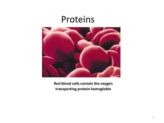
Proteins Red blood cells contain the oxygen transporting protein hemoglobin
Introduction • Proteins are essential parts of organisms and participate in every process within cells. • Many proteins are enzymes that catalyze biochemical reactions and are vital to metabolism. • Through the process of digestion, animals break down ingested protein into free amino acids that are then used in metabolism.
Aspects of a protein's structure • There are four distinct aspects of a protein's structure: • Primary structure, • Secondary structure, • Tertiary structure and • Quaternary structure.
Primary structure of proteins • This is the amino acid sequence • An amino acid exists as a dipolar salt • All amino acids have this general structure
Classification of amino acids • Non-polar with aliphatic R groups Glycine Alanine Proline Valine
Non-polar with aliphatic R groups Leucine Isoleucine Methionine
Aromatic R groups Phenylamine Tryptophan Tyrosine
Polar Uncharged R groups Serine Cysteine Threonine Asparagine Glutamine
Positively charged R groups Histidine Arginine Lysine
Negatively charged R groups Aspartate Glutamate
Formation of a peptide bond • Amino acids are linked by PEPTIDE BONDS which are covalent in nature • Peptide bond is an amide linkage formed by a condensation reaction (loss of water) • Brings together the alpha-carboxyl of one amino acid with the alpha-amino of another • Portion of the amino acid left in the peptide is termed the amino acid RESIDUE • Amino acids sometimes called RESIDUES • R groups remain UNCHANGED – remain active • N-terminal amino and C-terminal carboxyl are also available for further reaction
Amino acid residue • Definition of amino acid residue: • an amino acid molecule that has lost a water molecule by becoming joined to a molecule of another amino acid.
Formation of a peptide bond A peptide bond contains group
Example of a pentapeptide Ser-Gly-Tyr-Ala-Leu
Write the three-letter abbreviations for thefollowing tetrapeptide: Ala-Leu-Cys-Met **This is not the same as Met-Cys-Leu-Ala**
INSULIN • Insulin has 51 amino acids, divided between two chains. One of these, the A chain, has • 21 amino acids; the other, the B chain, has 30. The A and B chains are joined by disulfide • bonds between cysteine residues (Cys-Cys).
Secondary structure of proteins • This is the regularly repeating local structures stabilized by hydrogen bonds • Hydrogen bonds are electrostatic interactions between a donor consisting of the dipole of a polar O-H or N-H bond and an acceptor, consisting of an available lone pair of electrons on a neighbouring N or O atom. • Typical hydrogen bonds are about 5 - 10% as strong as a normal covalent bond, and are not permanent bonds like covalent bonds.
Hydrogen bonds in secondary structures of proteins • Although each hydrogen bond is relatively weak in isolation, the sum of the hydrogen bonds in a helix makes it quite stable. • The H-bonds result in a strong but temporary attraction between H-bonding partners.
An α -helical secondary structure. • Hydrogen bonds between ‘ backbone ’ amide NH and C= O groups stabilize the α -helix. • Hydrogen atoms of OH, NH or SH group (hydrogen donors) interact with free electrons of the acceptor atoms such as O, N or S
Hydrogen bonds in secondary structures of proteins : b-pleat
The parallel β -sheet secondary structure. • If the H-bonds are formed between peptide bonds in different chains, the chains become arrayed parallel or antiparallelto one another in what is commonly called a β -pleated sheet. • That is: When the zigzag polypeptide chains are arranged side by side, they form a structure resembling a series of pleats. • The β -pleated sheet is an extended structure as opposed to the coiled α -helix. • It is pleated because the carbon—carbon (C—C) bonds are tetrahedral and cannot exist in a planar configuration.
In an antiparallel arrangement, the successive β-strands alternate directions of the N and C-terminus. This is the most stable β-sheet arrangement. • In a parallel arrangement, the N-termini of successive strands are oriented in the same direction, generating a less stable β-sheet due to the non-planarity of the inter-strand H-bonds.
Tertiary Structure of Proteins • T he three-dimensional, folded and biologically active conformation of a protein is referred to as its tertiary structure. • This structure reflects the overall shape of the molecule. • T he three-dimensional tertiary structure of a protein is stabilized by interactions between side chain functional groups: covalent disulfide bonds, hydrogen bonds, salt bridges, and hydrophobic interactions.
Examples of amino acid side chain interactions contributing to tertiary Structure
Stabilizing interactions responsible for the tertiary structure of a protein.
Tertiary Structure of Proteins • These complex structures is held together by a combination of several molecular interactions that involve the R-groups of each amino acid in the chain. • These interactions include • hydrogen bonds between polar R- groups • ionic bonds between charged R-groups • hydrophobic interactions between non-polar R-groups • covalent bonds: The disulfide bond
Tertiary Structure of Proteins… • The importance of disulfide bonds in the structure of certain proteins is demonstrated by hair. Hair is made of the protein apha-keratin. The particular structure of your hair (straight, curly, etc.) is based on specific disulfide bonds that naturally form in the hair protein. This should help explain why an individual with straight hair cannot simply heat their hair, denature the protein (keratin), put in curlers and make it curly. The disulfide bonds are covalent bonds and thus are very strong. Heating these bonds will not break them, so simply heating hair will not change straight hair to curly.
Tertiary Structure of Proteins… • Instead, it is necessary to break these bonds chemically, reform the hair to the desired shape, and make new disulfide bonds to maintain the new shape. • If an individual goes to the hairdresser for a permanent, the beautician must first treat the hair with a reagent that reduces (and thus breaks) the disulfide bond, then put in curlers (to get the desired shape), and add an oxidizing agent to form new disulfide bonds to maintain the new shape.
Tertiary structure is important! The function of a protein (except as food) depends on its tertiary structure. If this is disrupted, the protein is said to be denaturedand it loses its activity. For example: • denatured enzymes lose their catalytic power • denatured antibodies can no longer bind antigen A mutation in the gene encoding a protein is a frequent cause of altered tertiary structure.
Quaternary structure of proteins • It is the shape or structure that results from the interaction of more than one protein molecule, usually called protein subunits, which function as part of the larger assembly or protein complex.
Examples of quaternary structures TetramerHexamerFilament SSBDNA helicaseRecombinase Allows coordinated Allows coordinated DNA binding Allows complete DNA binding and ATP hydrolysis coverage of an extended molecule
Some proteins (such as hemoglobin) have more than one peptide chain (these are multimeric proteins). The manner in which these chains fit together is the quaternary structure. • The subunits of a multimeric protein may be identical (homomultimericprotein), homologous or totally dissimilar (heteromultimeric protein ) and dedicated to disparate tasks. In some protein assemblies, one subunit may be referred to as a "regulatory subunit" and another as a "catalytic subunit." • The protein hemoglobin is made up of four polypeptide chains, two apha chains and two beta chains
The number of subunits in an oligomeric complex is described using names that end in –mer (Greek for "part, subunit") • 1 = monomer, 2 = dimer, 3 = trimer, 4 = tetramer, 5 = pentamer 6 = hexamer, 7 = heptamer, 8 = octamer, 9 = nonamer, 10 = decamer, 11 = undecamer, 12 = dodecamer etc • Although complexes higher than octamers are rarely observed for most proteins, there are some important exceptions: A capsid is the protein shell of a virus. It consists of several oligomeric (e.g. 60) structural subunits made of protein called protomers.
Major Classes of proteins Protein types • Proteins fall into three general classes, based on their overall three-dimensional (tertiary) structure and on their functional role: • fibrous, • membrane, • globular
Fibrous Proteins • Fibrous proteins tend to be long, narrow molecules. Fibrous proteins are used to construct macroscopic structures, especially structures outside of cells. Fibrous proteins tend to have a structural role, although some have more active functions as well. • Fibrous proteins are elongated molecules in which the secondary structure(either a-helices or b-pleated sheets) forms the dominant structure. • Fibrous proteins are insoluble, and play a structural or supportive role in the body, and are also involved in movement (as in muscle and ciliary proteins). • One feature of fibrous tissues is that they often have regular repeating structures.
Fibrous Proteins • Keratin, for example, which is found in hair, horns, wool, nails, and feathers, is a helix of helices (2 pairs of a-helices wound around one another) and has a seven amino acid repeating structure. Other keratins are found in skin, fur, hair, wool, claws, nails, hooves, horns, scales, beaks, feathers, actin and mysin in muscle tissues and fibrinogen needed for blood clots. • Fibroin is the fibrous protein that makes up silk cloth and spider webs. • Silk is a fibrous protein that is composed only of b-sheets. It too has a repeating pattern: layers of glycine alternate with layers of alanine and serines in the b-sheets. • Collagen, is the major protein component of connective tissue. In collagen, every third amino acid is glycine and many of the others are proline. • fibrous proteins generally have only primary and secondary structure whereas


