Download
1 / 57
610 likes | 1.08k Views
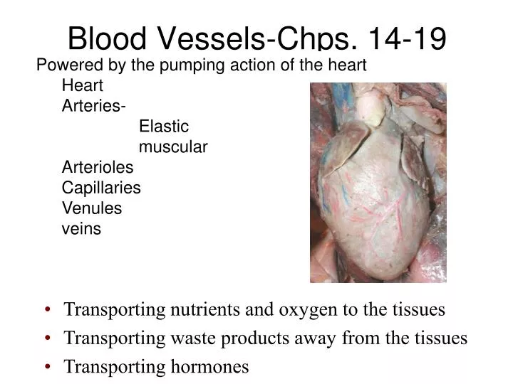
Powered by the pumping action of the heart Heart Arteries- Elastic muscular Arterioles Capillaries Venules veins. Blood Vessels-Chps. 14-19. Transporting nutrients and oxygen to the tissues Transporting waste products away from the tissues Transporting hormones.

E N D
Powered by the pumping action of the heart • Heart • Arteries- • Elastic • muscular • Arterioles • Capillaries • Venules • veins Blood Vessels-Chps. 14-19 • Transporting nutrients and oxygen to the tissues • Transporting waste products away from the tissues • Transporting hormones
I. Review anatomy of vessels A. Arteries B. Elastic C. Muscular D. Arterioles- resistance vessels E. Capillaries- exchange vessels F. Veins- capacitance vessels II. Ohm’s law is flow = change in pressure/ resistance A. Blood Flow i. Laminar vs. turbulent B. Pressure- blood pressure i. Mean arterial pressure (MAP) ii. Central venous pressure iii. Pulse pressure C. Resistance i. Factors of resistance- Poiseuille’s law III. Getting to know “Flow” better A. Velocity B. Control of flow i. Autoregulation ii. Nervous system iii. Endocrine-kidney (unit 4) Exchange of extracellular fluid- the microcirculation Starling Forces i. Capillary hydrostatic pressure ii. Interstitial hydrostatic pressure iii. Capillary colloid osmotic pressure iv. Interstitial colloid osmotic pressure Lymphatic drainage Causes of edema Lecture outline
Arteries • Branch and diverge • Blood away from heart • Walls have 3 tunics • Tunica intima-simple squamous endothelium • Tunica media-circular sheets of smooth muscle (vasodilation and vasoconstriction- diameter controlled by local factors and sympathetic NS) • Tunica adventitia- connective tissue with collagen and elastin in longitudinal arrangement
Arteries • Elastic- largest arteries near heart • Low resistance • More elastin interspersed with the tunica media • Can distend and recoil back to pump blood (maintain blood pressure) • Muscular- • Supply organs • Can regulate diameter of artery to control blood supply to organ • Thick tunica media with more smooth muscle • External and internal elastic lamina.
Arterioles • Smallest arteries- “resistance arteries” • THICK tunica media- little compliance • Diameter controlled by local factors (intrinsic) and sympathetic division (extrinsic) and long-term factors (hormones) • Metarterioles- just upstream of capillary beds. • Precapillary sphincters-controls blood reaching capillary bed.
Capillaries • Smallest blood vessels • Single layer of endothelial cells and basal lamina • Renew interstitial fluid- pick up wastes, drop off nutrients, etc. • Most cells only 20-30 µm away • Over 10 billion of them.
Types of Capillaries • Continuous • Most common and least permeable • Intercellular clefts and transcellular cytosis allows for exchange of molecules • Abundant in skin and muscle • Fenestrated • “Holes” in the endothelial membrane • Found in kidney • Sinusoidal/ discontinuous • Most permeable and least common • Big ‘holes” in endothelial membranes • Big clefts between cells • Liver, spleen, and bone marrow especially
Veins • Volume reservoir- “capacitance vessels” (60-70%) of blood • Have vasomotor control. • Valves in abdominal veins prevent backflow • Skeletal muscle “pump” and respiratory pump
100 ml Vascular Distensibility= is the fractional increase in volume for each mmHg rise in pressure times original volume- veins are 8x more distensible 0 mmHg 100 mmHg Artery Vein 800 ml In hemodynamics, it’s more valuable to know the total quantity of blood that can be stored in a given portion of the circulation for each mmHg pressure rise. Capacitance = increase in volume/increase in pressure The capacitance of veins is 24 times that of arteries.
Ohm’s Law • Q=P/R • Flow (Q) through a blood vessel is determined by: • 1) The pressure difference (P) between the two ends of the vessel • Directly related to flow • 2) Resistance (R) of the vessel • Inversely related to flow • Can you rearrange the equation above and solve for P? Solve for R?
Blood Flow (L/min) • Blood flowis the quantity of blood that passes a given point in the circulation in a given period of time. • Unit of blood flow is usually expressed as milliliters (ml) or Liters (L) per minute. • Overall flow in the circulation of an adult is 5 liters/min which is the cardiac output. • CO= HR X SV • 70 b/min x 70 ml/beat =4900ml/min
Characteristics of Blood Flow • Blood usually flows in streamlines with each layer of blood remaining the same distance from the wall, this type of flow is called laminar flow. • When laminar flow occurs, the velocity of blood in the center of the vessel is greater than that toward the outer edge creating a parabolic profile. Laminar flow Blood Vessel
Laminar Vs. Turbulent Blood Flow • Causes of turbulent blood flow: • high velocities • sharp turns in the circulation • rough surfaces in the circulation • rapid narrowing of blood vessels Turbulent flow • Laminar flow is silent, whereas turbulent flow tend to cause murmurs. • Murmurs or bruits are important in diagnosing vessels stenosis, vessel shunts, and cardiac valvular lesions.
Effect of Wall Stress on Blood Vessels Turbulent flow increases resistance and wall stress Nitric oxide released by endothelial cells to reduce the stress Aortic Aneurysm Atherosclerosis
Blood Pressure—The driving force Stephen Hales 1733 • Blood pressure (hydrostatic pressure) is the force exerted by the blood against any unit area of vessel wall. • Measured in millimeters of mercury (mmHg). A pressure of 100 mmHg means the force of blood was sufficient to push a column of mercury 100mm high. • All vessels have it – but we’re usually addressing arteries when we refer to it.
Ejected Blood contracted When the LV contracts more blood enters the arterial system than gets pushed onward. This causes the arteries to stretch and pressure within them to rise. The highest pressure achieved is known as the systolicpressure.
relaxed Recoil of the elastic artery As the LV relaxes, the stretched arterial walls recoil and push the contained blood onward through the system. As they recoil, the amount of blood contained decreases as does pressure. The lowest pressure achieved just before the next contraction is the diastolic pressure.
100 mmHg 100 mmHg A B 20 mmHg R = .1mmHg/ml/min R = .1mmHg/ml/min Mean Arterial Pressure (MAP) FLOW = arterial - venous pressure (P) resistance (R) • Is an average, but not a simple arithmetic average • Heart spends longer in diastole than systole • Value is significant- why? • The difference between the mean arterial pressure and the pressure in the venous system drives the blood through the capillary beds. • MAP= .4 (systolic) + .6 (diastolic)= 96mmHg • Venous pressure is about 2mmHg 0 mmHg FLOW = 1000 ml/min FLOW = 800 ml/min
Central Venous Pressure • Pressure in the right atrium is called central venous pressure. • determined by the balance of the heart pumping blood out of the right atrium and flow of blood from the large veins into the right atrium. • normally 0 mmHg, but can be as high as 20-30 mmHg. • More vigorous heart contraction (lower CVP). • Less heart contraction (higher CVP) • Factors that increase CVP: - increased blood volume - increased venous tone (peripheral pressure) - dilation of arterioles - decreased right ventricular function - Skeletal and respiratory pumps Figure 15-9; Guyton and Hall
Arterial Pulsations and Pulse Pressure • The height of the pressure pulse is the systolic pressure (120mmHg), while the lowest point is the diastolic pressure (80mmHg). • The difference between systolic and diastolic pressure is called the pulse pressure(40mmHg). Systolic Pressure } Pulse Pressure Diastolic Pressure
Factors Affecting Pulse Pressure • Stroke volume—increases in stroke volume increase pulse pressure, conversely decreases in stroke volume decrease pulse pressure. • Arterial compliance—decreases in compliance increases pulse pressure; increases in compliance decrease pulse pressure. Figure 15-5; Guyton and Hall
Heart rate Stroke volume Stroke volume Cardiac output Systolic Pressure } Pulse Pressure Mean Pressure Diastolic Pressure Arterial compliance Time Total Peripheral resistance HR x SV = CO = MAP/ TPR MAP= (0.4 SP) + (0.6 DP) PP= SP- DP
Damping of Pulse Pressures in the Peripheral Arteries What’s an anatomical reason for why the pressure fluctuation disappears here? • The intensity of pulsations becomes progressively less in the smaller arteries. • The degree of damping is proportional to the resistance of small vessels and arterioles and the compliance of the larger vessels. • Elastic arteries: • large radii, low resistance, some pressure reservoir • Muscular arteries • Smaller radii • Little more resistance • More pressure reservoir • Arterioles • Thick tunica media vs. radius • major pressure reservoir Figure 15-6; Guyton and Hall
Pulmonary veins Capillaries Arterioles Venules Blood Pressure Profile in the Circulatory System 1 2 0 1 0 0 8 0 Pressure (mmHg) Pulmonary arteries Venae cavae 6 0 Large veins Small veins Capillaries Venules 4 0 Large arteries Small arteries Arterioles 2 0 Aorta 0 Systemic Pulmonary • Circulatory pressure- averages 100mmHg • Arterial blood pressure-100-35mmHg • Capillary pressure- 35mmHg at beginning and 10-15mmHg at end • Venous pressure-15-0mmHg • Large pressure drop across the arteriolar-capillary junction
FLOW = P RESISTANCE Resistance • R = ΔP = mmHg • Q ml/min • Resistance is the impediment to blood flow in a vessel. • Can not be measured directly How Would a Decrease in Vascular Resistance Affect Blood Flow? FLOW = P RESISTANCE Conversely, Therefore, flow and resistance are inversely related!
Resistance makes a difference for the two sides of the heart! • Let’s say the CO (flow) is roughly 100ml/sec (easier math). • To calculate systemic resistance vs. pulmonary resistance we need to know pressure differences. • Pulmonary resistance is 16-2/100 • Systemic resistance is 100/100 • So, CO is same on each side of heart (has to be!), but right side generates less pressure due to lower resistance (1/7th than systemic). 100 mmHg 16 mmHg 2mmHg 0mmHg • R = ΔP = mmHg • Q ml/min
Factors of Resistance Poiseuille’s Law = Q =_Pr4 8l • Blood viscosity • Total vessel length • Vessel diameter • Resistance (length)(viscosity) • (radius)4
Viscosity • What are the major contributors to blood viscosity? • As viscosity increases, resistance will… • An increase in plasma EPO will cause resistance to… Figure 14-11; Guyton and Hall Figure 14-12; Guyton and Hall
Total Vessel Length • Longer the vessel.....more opportunity for resistance. Radius
Ohm’s Law Blood Flow (Q) = Δ P/ R Increase pressure- increase blood flow Decrease resistance- increase blood flow Increase resistance- decrease blood flow Vessel diameter Viscosity length Turbulence (usually result of an occlusion reducing vessel diameter unevenly) Blood flow in center is fastest- because that is the area of least resistance P1 P2 ΔP= P1-P2 So, lets review:Blood Flow is volume flowing/time • As resistance decreases, flow will… • As the pressure gradient increases, flow will… • Which does the heart influence more: pressure gradient or resistance?
Flow (amount of blood/time) MUST be the same through vessels in series! • If a pipe’s diameter changes over its length, a fluid will flow through narrower segments faster than it flows through wider segments because the volume of flow per second must be constant throughout the entire pipe. • Flow (volume/time) vs. velocity (distance/time) are NOT synonyms!
If capillaries have such a small diameter, why is the velocity of blood flow so slow? Aorta >Arterioles > Small veins >Capillaries We need slow blood flow in the capillaries—the exchange vessels
Control of blood flow through vessels- Why is this important? • Perfusion vs. ischemia vs. hypoxia vs. anoxia vs. infarction • Tissue Perfusion Dependent on: • Cardiac output • Peripheral resistance • Blood pressure • Regulation of perfusion dependent on: • Autoregulation (Acute, local, intrinsic) • Neural mechanisms (acute) • Endocrine mechanisms (long-term) http://www.flometrics.com/services/artery/
Autoregulation the automatic adjustment of blood flow to each tissue in proportion to the tissue’s requirements at any instant even over a wide range of arterial pressures Working Muscle Tissue active hyperemia: when tissues become active, blood flow increases. Aka: intrinsic metabolic vasodilation Tissue temp. rises Arterioles serving tissue vasodilate and precapillary sphincters relax Tissue CO2 levels rise Tissue O2 levels fall Lactic acid levels rise Increased blood flow to tissue CO2 removed Now arterioles will vasoconstrict and precapillary sphincters contract Lactic acid removed Heat removed O2 delivered
Autoregulation of Blood Flow to specific tissues • Vasodilator agents Histamine Nitric oxide Elevated temperatures Potassium/hydrogen ions Lactic acid Carbon dioxide Adenosine/ ADP • Vasoconstrictors Norepinephrine and epinephrine Angiotensin Vasopressin (ADH) Thromboxane
Other ways to ultimately change blood flow throughout the body is to change Pressure and Resistance Arterial Pressure = Cardiac Output x Total Peripheral Resistance Short term BP control- nervous Long Term BP control- hormonal
Brain Centers involved in Short Term BP Control • Vasomotor • Adjusts peripheral resistance by adjusting sympathetic output to the arterioles • Cardioinhibitory- transmits signals via vagus nerve to heart to decrease heart rate. (parasympathetic) • Cardioacceleratory/ contractility-sympathetic output
Vasomotor control: Sympathetic Innervationof Blood Vessels • Sympathetic nerve fibers innervate all vessels except capillaries and precapillary sphincters (precapillarysphincters follow local control) • Innervation of small arteries and arterioles allow sympathetic nerves to increase vascular resistance. • Large veins and the heart are also sympathetically innervated. Figure 18-2; Guyton and Hall
Anatomy of the Baroreceptors • spray type nerve endings located in the walls of the carotid bifurcation called the carotid sinus and in the walls of the aortic arch-pressoreceptors that respond to stretch. • Signals from the carotid sinus are transmitted by the glossopharyngeal nerves . • Signals from the arch of the aorta are transmitted through the vagus into the NTS. • Important in short term regulation of arterial pressure. • They are unimportant in long term control of arterial pressure because the baroreceptors adapt. Figure 18-5; Guyton and Hall
Arterial Pressure Response of the Baroreceptors to Arterial Pressure Figure 18-7; Guyton and Hall Constrict Common Carotids • Baroreceptors respond to changes in arterial pressure. • As pressure increases the number of impulses from carotid sinus increases which results in: 1) inhibition of the vasoconstrictor 2) activation of the vagal center Pressure at Carotid Sinuses Constrictors Figure 18-5; Guyton and Hall
Functions of the Baroreceptors • Maintains relatively constant pressure despite changes in body posture. Decrease Venous return Supine Standing Sympathetic Nervous Activity Decrease Cardiac Output Vasomotor Center Sensed By Baroreceptors Decrease Arterial Pressure
BP rises Decreased vasomotor activity Decreased NE release on arterioles Vasodilation Detected by baroreceptors in aortic arch & carotid sinus Decreased PR Increased cardioinhibitory activity Info sent to cardiac and vasomotor centers Increased vagus activity Decreased BP Increased ACh release on heart Decreased cardioacceleratory activity Decreased CO Decreased NE release on heart Decreased SV and HR
Carotid and Aortic Chemoreceptors • Chemoreceptors are chemosensitive cells sensitive to oxygen lack, CO2 excess, or H ion excess. • Chemoreceptors are located in carotid bodies near the carotid bifurcationand on the arch of the aorta. • Activation of chemosensitive receptors results in excitation of the vasomotor center. Figure 18-5; Guyton and Hall O2 CO2 pH Chemoreceptors VMC Sympathetic activity BP
Vasomotor Center Heart rate Contractility Vagal Atrial Stretch afferents Nervous control also found in the heart- Bainbridge Reflex • Prevents damming of blood in veins, atria and pulmonary circulation. • Increase in atrial pressure increases heart rate. • Stretch of atria sends signals to VMC via vagal afferents to increase heart rate and contractility.
The Microcirculation-chapter 16 • Important in the transport of nutrients to tissues. • Site of waste product removal. • Over 10 billion capillaries with surface area of 500-700 square meters perform function of solute and fluid exchange. Figure 16-1; Guyton and Hall
Most substances are exchanged via diffusion • Concentration differences across capillary enhances diffusion.
Determinants of Net Fluid Movement across Capillaries-Starling forces • Capillary hydrostatic pressure (Pc)-tends to force fluid outward through the capillary membrane. (30 mmHg arterial; 10mmHg venous- average 17.3mmHg) • Interstitial fluid hydrostatic pressure (Pif)- opposes filtration when value is positive (but it’s not positive-- due to lymphatic drainage! – 3mmHg). Figure 16-5; Guyton and Hall
Determinants of Net Fluid Movement across Capillaries-Starling forces • Plasma colloid osmotic pressure ( c)- opposes filtration causing osmosis of water inward through the membrane • Colloid osmotic pressure of the blood plasma. (28mmHg) • 75% from albumin; 25% from globulins • Interstitial fluid colloid pressure ( if) promotes filtration by causing osmosis of fluid outward through the membrane • Colloid osmotic pressure of the interstitial fluid. (8mmHg) • 3gm% Figure 16-5; Guyton and Hall
Net Forces in Capillaries Filtration= Kf X (Pc- Pif - c + if) mmHg Mean forces tending to move fluid outward: Mean Capillary pressure 17.3 Negative interstitial free fluid pressure 3.0 Interstitial fluid colloid osmotic pressure 8.0 TOTAL OUTWARD FORCE 28.3 Mean force tending to move fluid inward: Plasma colloid osmotic pressure 28.0 TOTAL INWARD FORCE 28.0 Summation of mean forces: Outward 28.3 Inward 28.0 NET OUTWARD FORCE 0.3 Net filtration pressure of .3 mmHg which causes a net filtration rate of 2ml/min for entire body (2-4 liters/day!)