Download

1 / 15
150 likes | 677 Views
Anatomy of the Bladder & Urethra. Dr. Nimir Dr. Safaa. Objectives Describe the normal site, size, shape, and position of urinary bladder. Delineate the borders and surfaces of urinary bladder. Discuss the blood and nerve supply of the urinary bladder

E N D
Anatomy of the Bladder & Urethra Dr. Nimir Dr. Safaa
Objectives • Describe the normal site, size, shape, and position of urinary bladder. • Delineate the borders and surfaces of urinary bladder. • Discuss the blood and nerve supply of the urinary bladder • Discuss the lymphatic drainage of urinary bladder. • Describe the normal site, shape and length of male and female urethra. • Discuss urethra relations, blood and nerve supply, and their lymphatic drainage. • Describe thesphincters of urethra.
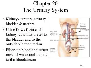
The Urinary Bladder Body Apex Fundus • It is a hollow muscular organ • It is a pelvic organ after puberty • It lies behind and superior to pubic bones leaving retropubic space in between • It is divided into apex, fundus and body • It has 4 surfaces, superior, posterior (base) and two inferiolateral • It is freely movable except at the neck that is attached by lateral ligaments of bladder and puboprostatic ligament in male andpubovesical ligament in female
The Urinary Bladder Cont., Posterior relation in male: • Vas deferens • Seminal vesicle • Rectum • Rectovesical fascia • Peritoneum Posterior relation in female: • Vagina and part of uterus Superior relation in male: • Peritoneum • Coils of ileum • Sigmoid colon Superior relation in female: • Uterus Lateral relation • Obturatorinternus muscle • Levatorani muscle
The Urinary Bladder Cont., Anterior relation • Symphysis pubis • Retropubic fat Inferior relation: • Prostate gland • The muscle of the bladder wall is called Detrusor muscle • It is thickened at the neck to form involuntary internal urethral sphincter • Trigone is triangular area where the two ureters and urethra open into its angles
Blood Supply,venous & lymphatic drainage of the bladder: • Superior vesical artery • Inferior vesical artery • Vaginal artery replace inferior vesiacal artery in female • In male, venous plexus around the bladder and prostate drain into inferior vesical vein • Also, superior vesical vein drains the bladder • Both veins drain into internal iliac vein • In female, venous plexus around bladder drain into vaginal or uterovaginal vein and then to internal iliac vein • Also, superior vesicalvein drains the bladder. • The lymphatic vessels pass mainly to the internal iliac lymph nodes; a few vessels drain into the external iliac lymph nodes
Nerve Supply: • The sympathetic fibers originate in the first and second lumbar ganglia and descend to the bladder via the hypogastric plexuses. The parasympathetic fibers arise as pelvic splanchnic nerves from the second, third, and fourth sacral nerves; they pass through the inferior hypogastric plexuses to reach the bladder wall. Most afferent sensory fibers of the bladder reach the central nervous system via the pelvic splanchnic nerves. Some afferent fibers travel with the sympathetic nerves. • Parasympathetic fibers leads to contraction of smooth fibers in ureter and bladder and relaxation of smooth fibers in internal urethric sphincter • Sympathetic fibers cause the opposite effect
Male Urethra • It is 20 cm long in male from the bladder neck to external urethral meatus • It has a tortous course • It divides into three parts: 1. Prostatic part: widest portion It is 3 cm long Most dilatable part Ducts of prostate gland and ejaculatory ducts open in it 2. Membranous part: Lies within urogenital diaphragm It is 1.5 cm long Least dilatable part
Male Urethra cont., 3. Penile part: It is 15.5 cm Is surrounded by erectile tissue of bulb and corpus spongiosum It is dilated at the end as navicular fossa Bulbourethral and penile glands open in it
Female Urethra • It is 4 cm in length and 6 mm in diameter • Extends from neck of bladder to external meatus • It passes under the symphysis pubis • It lies anterior to vagina • It opens in the vestibule anterior to vaginal opening • Paraurethral glands They are mucus secreting glands located at the sides of external meatus • It is easily dilatable • It is straight
Blood Supply of Urethra Male Urethra Female Urethra • Prostatic branches of inferior vesical and middle rectal arteries • Dorsal artery of the penis • Arteries of the bulb of the penis • Internal pudendal artery • Vaginal artery
Nerve Supply of Urethra • Parasympathetic supply From pelvic plexus made of S2-4 roots to form pelvic splanchnic nerve • Sympathetic supply T12-L1-2 Form hypogastric plexus and then form pelvic • Somatic supply Pudendal nerve from sacral plexus Sensory and motor to external urethral sphincter