Download
1 / 33
330 likes | 406 Views
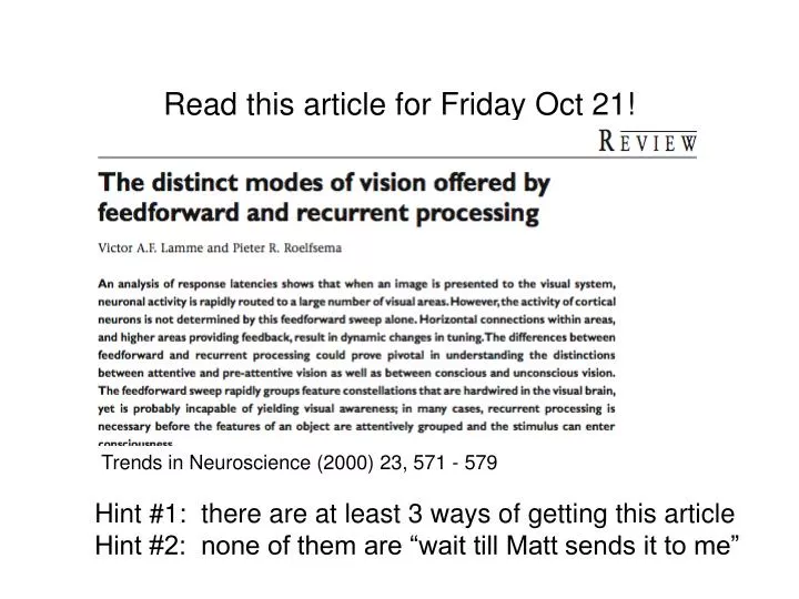
Read this article for Friday Oct 21!. Trends in Neuroscience (2000) 23, 571 - 579. Hint #1: there are at least 3 ways of getting this article Hint #2: none of them are “wait till Matt sends it to me”. Visual Pathways. Retina has distinct layers. Visual Pathways.

E N D
Read this article for Friday Oct 21! Trends in Neuroscience (2000) 23, 571 - 579 Hint #1: there are at least 3 ways of getting this article Hint #2: none of them are “wait till Matt sends it to me”
Visual Pathways • Retina has distinct layers
Visual Pathways • Retina has distinct layers • Amacrineand bipolar cells perform “early” processing • Peripheral retina features convergence of receptors onto ganglion cells • Foveal retina features divergence • Why!?
Visual Pathways • Retina has distinct layers • Amacrineand bipolar cells perform “early” processing • lateral inhibition at middle layer leads to centre-surround receptive fields • first step in shaping “tuning properties” of higher-level neurons
Visual Pathways • Retina has distinct layers • Ganglion cells project mainly to Lateral Geniculate Nucleus (LGN) of the thalamus • two kinds of ganglion cells: Magnocellular and Parvocellular • Magno is myelinated • Parvo is not myelinated • In what way does this matter? • visual information is already being shunted through functionally distinct pathways
Visual Pathways • visual hemifields project contralaterally • exception: bilateral representation of fovea! • Optic nerve splits at optic chiasm • about 90 % of fibers project to cortex via LGN • about 10 % project through superior colliculus and pulvinar • but that’s still a lot of fibers! Note: this will be important when we talk about visuospatial attention
Visual Pathways • Lateral Geniculate Nucleus maintains segregation: • of M and P cells (mango and parvo) • of left and right eyes P cells project to layers 3 - 6 M cells project to layers 1 and 2
Visual Pathways • Cortical regions vary in their anatomical connections and their functional specialization • Number should be thought of very loosely as “consecutive” as in a processing hierarchy • But note that this view is outdated as we’ll discover in reading the article by Lamme
Visual Pathways • Primary visual cortex receives input from LGN • also known as “striate” because it appears striped when labeled with some dyes • also known as V1 • also known as Brodmann Area 17
Visual Pathways • Primary cortex maintains distinct pathways – functional segregation • M and P pathways synapse in different layers • Ascending (i.e. feed-forward) projections synapse in middle layers • Descending (i.e. feed-back) projections synapse in superfical and deep layers W. W. Norton
Visual Pathways • Visual scene is represented: • Retinotopically thus… • spatiotopically = Fovea Tootell R B H et al. PNAS 1998;95:811-817
How does the visual system represent visual information? How does the visual system represent features of scenes? • Vision is analytical - the system breaks down the scene into distinct kinds of features and represents them in functionally segregated pathways
Visual Neuron Responses • The notion of a receptive field is fundamental in vision science • A neuron’s receptive field is the region in space in which a stimulus will evoke a response from that neuron • Receptive field properties vary widely across visual neurons and are never just “ON” or “OFF” • Unit recordings in LGN reveal a centre/surround receptive field
Visual Neuron Responses • Unit recordings in LGN reveal a centre/surround receptive field • many arrangements exist, but the “classical” RF has an excitatory centre and an inhibitory surround • these receptive fields tend to be circular - they are not orientation specific How could the outputs of such cells be transformed into a cell with orientation specificity?
Visual Neuron Responses • LGN cells converge on “simple” cells in V1 imparting orientation (and location) specificity
Visual Neuron Responses • LGN cells converge on “simple” cells in V1 imparting orientation (and location) specificity • Again, information is physically seperated into a “map”
Visual Neuron Responses • LGN cells converge on simple cells in V1 imparting orientation specificity • Thus we begin to see how a simple representation – orientations of lines - can be maintained in the visual system • increase in spike rate of specific neurons indicates presence of a line with a specific orientation at a specific location on the retina • Reality is that spike rate probably is only one part of the story: information is coded in many ways e.g. • Relative timing • Graded potentials
The Role of “Extrastriate” Areas • Different visual cortex regions contain cells with different tuning properties
The Role of “Extrastriate” Areas • Consider two plausible models: • System is hierarchical: • each area performs some elaboration on the input it is given and then passes on that elaboration as input to the next “higher” area • System is analytic and parallel: • different areas elaborate on different features of the input
The Role of “Extrastriate” Areas • Functional imaging (PET) investigations of motion and colour selective visual cortical areas • Zeki et al. • Subtractive Logic • stimulus alternates between two scenes that differ only in the feature of interest (i.e. colour, motion, etc.)
The Role of “Extrastriate” Areas • Identifying colour sensitive regions Subtract Voxel intensities during these scans… …from voxel intensities during these scans …etc. Time ->
The Role of “Extrastriate” Areas • result • voxels are identified that are preferentially selective for colour • these tend to cluster in anterior/inferior occipital lobe
The Role of “Extrastriate” Areas • similar logic was used to find motion-selective areas Subtract Voxel intensities during these scans… …from voxel intensities during these scans …etc. STATIONARY STATIONARY MOVING MOVING Time ->
The Role of “Extrastriate” Areas • result • voxels are identified that are preferentially selective for motion • these tend to cluster in superior/dorsal occipital lobe near TemporoParietal Junction • Akin to Human V5
The Role of “Extrastriate” Areas • Thus PET studies doubly-dissociate colour and motion sensitive regions
The Role of “Extrastriate” Areas • V4 and V5 are doubly-dissociated in lesion literature:
The Role of “Extrastriate” Areas • V4 and V5 are doubly-dissociated in lesion literature: • achromatopsia (color blindness): • there are many forms of color blindness • cortical achromatopsia arises from lesions in the area of V4 • singly dissociable from motion perception deficit - patients with V4 lesions have other visual problems, but motion perception is substantially spared
The Role of “Extrastriate” Areas • V4 and V5 are doubly-dissociated in lesion literature: • akinetopsia (motion blindness): • bilateral lesions to area V5 (extremely rare) • severe impairment in judging direction and velocity of motion - especially with fast-moving stimuli • visual world appeared to progress in still frames • similar effects occur when M-cell layers in LGN are lesioned in monkeys
Visual Neuron Responses • Edges are important because they are the boundaries between objects and the background or objects and other objects
Visual Neuron Responses • This conceptualization of the visual system was “static” - it did not take into account the possibility that visual cells might change their response selectivity over time • Logic went like this: if the cell is firing, its preferred line/edge must be present and… • if the preferred line/edge is present, the cell must be firing • We will encounter examples in which these don’t apply! • Representing boundaries must be more complicated than simple edge detection!
Visual Neuron Responses • Boundaries between objects can be defined by color rather than brightness
Visual Neuron Responses • Boundaries between objects can be defined by texture
Visual Neuron Responses • Boundaries between objects can be defined by motion and depth cues