Download
1 / 62
620 likes | 651 Views
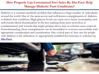
Explore the intricate anatomy of the foot, its ligaments, muscles, nerves, and blood supply. Learn about foot kinetics and kinematics, gait cycles, and injury prevention techniques through physical conditioning and proper footwear. Discover common toe and foot conditions, including deformities like pes cavus and pes planus, along with management strategies.

E N D
Foot Conditions Chapter 19
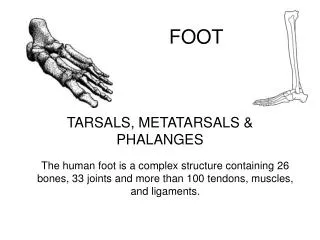
Anatomy (Cont’d) • Forefoot • Metatarsals and phalanges; numerous joints • Support and distribute body weight throughout the foot
Anatomy (cont.) • Midfoot • Navicular, cuboid, 3 cuneiforms; numerous joints • Talocalcaneonavicular joint (TCN) • Talus moves simultaneously on calcaneus and navicular • Combined action of talonavicular and subtalar joint • Close-packed position—supination
Ligaments supporting the midfoot and hindfoot region Anatomy (cont.)
Anatomy (cont.) • Hindfoot • Calcaneus and talus • Talocrural joint (ankle joint) • Articulation of talus, tibia, and fibula • Close-packed position—dorsiflexion • Medial ligament—deltoid • Lateral ligament—anterior talofibular; posterior talofibular; calcaneofibular
Hindfoot Subtalar joint Behaves as a flexible structure Axis of rotation of the subtalar joint lies oblique in the sagittal and frontal planes Anatomy (cont.)
Anatomy (cont.) • Plantar arches • Support and distribute body weight • Longitudinal arch— medial and lateral • Transverse arch • Ligaments • Spring (calcaneonavicular) • Long plantar • Short plantar
Anatomy (cont.) • Plantar arches • Plantar fascia
Anatomy (cont.) • Muscles • Lateral and medial view
Anatomy (cont.) • Muscles • Posterior view
Anatomy (cont.) • Muscles • Intrinsic muscles of the foot – dorsal view
Anatomy (cont.) • Muscles • Intrinsic muscles of the foot – plantar view
Anatomy (cont.) • Nerves • Sciatic nerve • Tibial nerve • Common peroneal nerve — deep and superficial peroneal nerves • Femoral — saphenous
Anatomy (cont.) • Blood supply • Femoral artery • Popliteal • Anterior and posterior tibial • Anterior tibial • Dorsal pedal
Kinematics • Gait cycle • Consists of alternating periods of single-leg and double-leg support • Requires a set of coordinated, sequential joint actions of the lower extremity
Kinematics (cont.) • Motions • Toe — flexion and extension • Ankle (subtalar) — dorsiflexion and plantarflexion • Foot and ankle • Inversion and eversion • Pronation and supination
Kinetics • Bones subject to several loading patterns • Running • Foot sustains forces 2–3× body weight • Bones are typically 2–4× strength needed • Repeated forces—stress fractures • Foot deforms during weight bearing • Absorbing a smaller force of longer duration than if it were rigid • Deformation causes storage of mechanical energy in the stretched tendons, ligaments, and plantar fascia
Injury Prevention • Physical conditioning • Strengthening • Extrinsic muscles • Intrinsic muscles • Flexibility • Achilles tendon • Footwear • Demands of sport; wear shoe for its intended purpose • Proper fit • Protective equipment • Taping; braces; orthotics
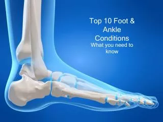
Toe and Foot Conditions • Foot Deformities • Pes cavus • High arch and rigid foot • Pes planus • Flatfoot and mobile foot • Associated with common injuries (refer to Box 19.1)
Toe and Foot Conditions (cont.) • Toe deformities • Hallux rigidis • Degenerative arthritis in first MTP • S&S • Tender, enlarged first MTP joint • Loss of motion • Difficulty wearing shoes with an elevated heel • Hallmark sign—restricted toe extension • Management: shoe modification
Toe and Foot Conditions (cont.) • Toe deformities • Hallux valgus • Thickening of the medial capsule and bursa, resulting in severe valgus deformity of great toe • Asymptomatic or symptomatic • Treatment—symptomatic
Toe and Foot Conditions (cont.) • Hammer toe • Extension of MTP joint, flexion at PIP joint, and hyperextended at the DIP joint • Claw toe • Hyperextension of MTP joint and flexion of DIP and PIP joints • Mallet toe • Neutral position at MTP and PIP joints, flexion at DIP joint • Difficult to treat conservatively
Toe and Foot Conditions (cont.) • Turf toe • Sprain of the plantar capsular ligament of 1st MTP joint • Mechanism: forced hyperflexion or hyperextension of great toe • Acute or repetitive overload • Valgus ↑ susceptibility • S&S • Pain, point tenderness, and swelling on plantar aspect of MP joint • Extreme pain with extension • Potential for tear in flexor tendons or fracture of sesamoid bones • Management: standard acute; rest; protection from excessive motion
Toe and Foot Conditions (cont.) • Ingrown toenail • Preventable with proper hygiene and nail care • Edge of nail grows into lateral nail fold and surrounding skin • Nail margin reddens; painful • Paronychia—fungal or bacterial infection • Management: refer to Application Strategy 19.2
Toe and Foot Conditions (cont.) • Metatarsalgia • General discomfort around the metatarsal heads • Constant overloading leads to flattening of transverse arch • Contributing factors—intrinsic and extrinsic (refer to Box 19.2) • Management: activity modification; footwear examination; strengthening exercises
Toe and Foot Conditions (cont.) • Bunion • Medial aspect of MTP joint of great toe; lateral aspect of the 5th toe • Thickening of capsule and bursa • Due to constant rubbing against inside of shoe • S&S (as condition worsens) • Lateral shift of great toe • Rigid, nonfunctional hallux valgus deformity • Once deformity occurs, little can be done to correct condition
Toe and Foot Conditions (cont.) • Retrocalcaneal bursitis • Due to external pressure—constrictive heel cup, coupled with excessive pronation or varus hindfoot • “Pump bump” • Management: standard acute; shoe modification; AT stretching
Foot Contusions • Trauma to the midfoot or forefoot: need to rule out fracture and damage to extensor tendons • Hindfoot—heel bruise • Thick padding of adipose tissue—does not always suffice • Stress in running, jumping, changing directions • S&S • Severe pain in heel • Unable to bear weight • Management: cold; heel cup or doughnut pad • Condition may persist for months
Toe and Foot Sprains • IP & MP joints • Sprains of MP and IP joints of the toes may occur by tripping or stubbing the toe • S&S • Pain, dysfunction, immediate swelling • Dislocation—gross deformity • Management—strapping
Toe and Foot Sprains (cont.) • Midfoot sprains • Mechanism: severe dorsiflexion, plantarflexion, or pronation • More frequent in activities in which the foot is unsupported • S&S • Pain and swelling is deep on medial aspect of foot • Weight bearing may be too painful • Management: standard acute; limited weight bearing
Overuse Conditions • Plantar fasciitis • Extrinsic and intrinsic risk factors • S&S • Pain with first steps in the morning • Point tenderness at medial calcaneal tubercle • ↑ pain with passive extension of great toe and ankle dorsiflexion • ↑ pain with weight bearing • Pain relieved with activity, but recurs after rest • Management: standard acute; refer to Application Strategy 19.4
Neurologic Conditions • Plantar interdigital neuroma (Morton’s neuroma) • Trauma or repetitive stress → abnormal pressure on plantar digital nerves • Common—web space between 3rd and 4th metatarsals; less common, between 2nd and 3rd metatarsals
Neurologic Conditions (cont.) • S&S • Sensation of having a stone in the shoe that worsens when standing • Tingling or burning, radiating to the toes, along with intermittent symptoms of a sharp shock-like sensation • Pain subsides when activity is stopped or when the shoe is removed; desire to remove the shoe and massage foot—classic sign • Management: metatarsal pad; broad, soft-soled shoe with a low heel
Neurologic Conditions (cont.) • Tarsal tunnel syndrome • Posterior tibial nerve (or branch) constricted beneath fibrous roof of foot flexor retinaculum • Often linked to excessive pronation or excessive valgus deformity • S&S • Pain at medial malleolus radiating into sole and heel • Paresthesia, dysesthesia, or hyperesthesia in nerve distribution • + Tinel’s sign • Management: rest; NSAIDs; orthoses; gradual return to activity
Foot and Lower Leg Fractures • Repetitive microtraumas → apophyseal or stress fractures • Tensile forces associated with severe ankle sprains → avulsion fractures of 5th metatarsal • Severe twisting → displaced and undisplaced fractures in foot, ankle, or lower leg
Foot and Lower Leg Fractures (cont.) • Freiberg's disease • Avascular necrosis of 2nd metatarsal head • Active adolescents ages 14–18 • Sever's disease • Traction-type injury of calcaneal apophysis • Seen in ages 7–10 • S&S • Heel pain with activity • + “squeeze” test • + Sever’s sign • Decreased heel cord flexibility • Management: standard acute; physician referral
Foot and Lower Leg Fractures (cont.) • Stress fractures • Often seen in running and jumping, especially after significant ↑ training mileage; change in surface, intensity, or shoe type • Common sites • 2nd metatarsal • Sesamoid bones • Navicular • Calcaneus • Tibia and fibula
Foot and Lower Leg Fractures (cont.) • S&S • Pain begins insidiously; ↑ with activity and ↓ with rest • Pain usually limited to fracture site • Pain with percussion, tuning fork, or ultrasound • Management: standard acute; physician referral
Foot and Lower Leg Fractures (cont.) • Avulsion fractures • Eversion sprain—deltoid ligament avulses portion of distal medial malleolus • Inversion sprain—plantar aponeurosis or peroneus brevis tendon avulses base of 5th metatarsal (type II) • Jones fracture • Type I transverse fracture into the proximal shaft of 5th metatarsal at junction of diaphysis and metaphysis • Often overlooked in conjunction with a severe ankle sprain • Complications: nonunions and delayed unions are common • Management: standard acute; physician referral
Phalanges/ metacarpals Standard S&S Relatively minor Tarsal fractures LisFranc injury Disruption of tarsometatarsal joint, with or without associated fracture Caused by a severe twisting injury Foot and Lower Leg Fractures (cont.)
Foot and Lower Leg Fractures (cont.) • 1st metatarsal dislocated from 1st cuneiform; other 4 metatarsals are displaced laterally, usually in combination with fracture at base of 2nd metatarsal • History of severe midfoot pain, paresthesia, or swelling in midfoot region with variable flattening of arch or forefoot abduction
Foot and Lower Leg Fractures (cont.) • Lateral process of talus • Due to traumatic ankle sprain • Persistent ankle pain; inability to walk for long periods • Posterior fracture to talus • Forced plantarflexion • Pain with running, jumping; resisted plantarflexion and great toe flexion • Neck of talus • Forced dorsiflexion • May compromise blood supply to talus