Download
1 / 37
370 likes | 439 Views
Learn about antibodies, their synthesis, and applications in bioseparation and immunoassays. Discover how antibodies are synthesized in vivo and utilized in affinity chromatography and ELISA. Explore the genetic mechanisms diversifying antigen repertoires and antibody production via hybridoma cells. Delve into the immunoresponse, antibody therapy, and drug delivery. Gain insights into the specific detection and separation processes in bioprocesses like affinity chromatography. Uncover the principles of immunoassays, including RIA, EIA, and LIA, and the techniques and methods crucial in enzyme immunoassays.

E N D
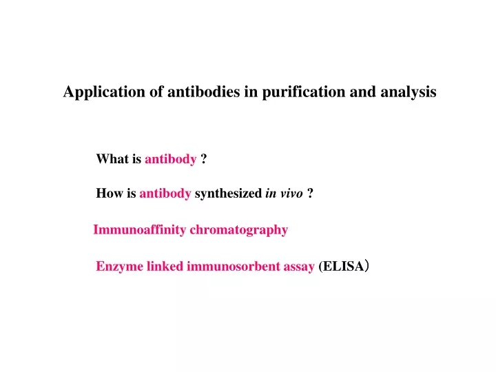
Application of antibodies in purification and analysis What is antibody ? How is antibody synthesized in vivo ? Immunoaffinity chromatography Enzyme linked immunosorbent assay (ELISA)
(Variable region) Light chain (Constant region) Heavy chain What is antibody ? Antibody (IgG type) VH VL CH1 CL CH2 CH3 Protein secreted from lymphocytes (B cell, plasma cell) Several classes, IgG MW about 150000 Binding with many kinds antigens (proteins, sugars, toxins, virus, microorganisms and so on)
(Variable region) Light chain (Constant region) Heavy chain Antibody (IgG type) VH VL CH1 CL CH2 CH3 Variable region Antigen binding site ------- Binding with wide variety of antigens Complementarity-determining region, CDR Constant region Opsonization, Compliment activation, Activation of NK cell Large number of repertoire and specificity against antigens
Why antibodies can bind with wide variety of antigens ? Huge number of repertory Hypervariable regions gene variation CDR regions in VH and VL Rearrangement of gene in B and T cells What is the genetic mechanism of diversification of antigen repertoire ?
antibodies Clonal selection of B cell repertoire by antigen
Preparation of hybridoma cells producing a monoclonal antibody • Monoclonal antibody • Homogeneous antibody that recognizes a single type of antigenic site 1. B cells from an immunized animal are fused with immortal lymphocytes called myeloma cells. 2. In HAT medium, the unfused cells and the myeloma cells do not grow. The B cells have a limited life span in culture. Only fused cells formed from a myeloma cell and a B cell (hybridoma cells) can survive in HAT medium. 3. Each selected hybridoma cell is tested for the production of the desired antibody. (containing hypoxanthine, aminopterin, and thymidine)
How is antibody synthesized in vivo ? Immunosystem The body is protected from infectious agents and from other harmful substances. Humoral immunity Immunity mediated by antibodies Neutralization of toxin, marking of bacterial cells and virus Cell-mediated immunity T lymphocytes Killing infected cells Activate to effector cells
Application of antibodies Drug delivery, antibody therapy Neutralization of toxin, marking of bacterial cells and virus Targeting therapy Immunoassay Specific detection from complicated mixture
Bioseparation (affinity chromatography) Specific adsorption washing Elution High purity separation in bioprocesses
a-amylase SDS-PAGE of secreted of a-amylase in supernatant
Purified a-amylase Crude broth
Difference in molecules × binding non binding Assay of biological materials by use of antigen (Immunoassay) • Advantages of immunoassay • Specificity and high affinity • --- high sensitivity assay from mixture • Recognition subtle difference in similar molecules
Immunoassay Radioimmunoassay (RIA) Enzyme immunoassay (EIA) homogeneous enzyme immunoassay heterogeneous enzyme immunoassay (Enzyme linked immunosorbent assay (ELISA)) Liposome immunoassay (LIA) Flow-injection analysis
Classification of enzyme immunoassay Solid phasecompetitivetwo antibody solid phase method a) non-competitivedirect method b) sandwich method c) forward sandwich method simultaneous two site method liposome immunoassay Homogeneouscompetitive enzyme multiple immunoassay technique d) non-competitive liposome immune lysis assay
Antigen in sample Enzyme labeled antigen E E E E Enzyme labeled antibody Adsorbed antibody Enzyme labeled hapten (a) (b) (d) (c) Schemes of enzyme immunoassay
E Enzyme-conjugated antibody Relation between signal intensity and concentration
Enzyme labeled antigen E Signal, arbitrary unit Antigen in sample Relation between signal and sample concentration in competitive methods Signal from enzyme labeled antigen Amount of sample antigen coupled to antibody
* Enzyme immunoassay Enzyme linked immunosorbent assay (ELISA) Liposome immunosorbent assay (LISA) Color signal depended on antigen concentration
Enzyme Immunoassay Enzyme conjugated antibody against target antigen Enzyme-substrate reaction Washing Substrate
Antigen Blocking of surface Blocking agent Adsorption of primary antibody Primary antibody Adsorption of labeled antibody Enzyme-conjugate antibody Color development Adsorption of antigen ELISA
Washing Washing Immobilization of ligand protein Binding analyte Blocking Solid-liquid reaction B/F separation Washing Washing Long time consuming Multiple steps for detection Enzyme-conjugated antibody Detection Operation in sandwich ELISA
Liposome Immunoassay Enzyme Immunoassay Enzyme-conjugated antibody Immunoliposome Few enzyme molecules conjugated with one molecule of antibody Large number of enzyme molecules encapsulated in a liposome
Diameter of liposome ▲350 nm ▲200 nm ▲130 nm ▲60 nm ▲ELISA Coating antigen, human IgM concn, 10 mg/ml Immunoliposome, anti-rabbit IgG (goat) concn, 3mg/ml Merits of immunoliposome High signal intensity Low signal High signal
* Liposome immunosorbent assay (LISA) Advantages of LISA high sensitivity high level of signal * Membrane blotting assay *automated micro-assay method labeled antibody (conventional) *limited sensitivity and strength of signal Blotting assay using immunoliposomes
HRP Immunoliposome Reaction products precipitated Substrate (4-chloro-1-naphthol) Primary antibody Antigen Membrane HRP-conjugated antibody Membrane conventional immunoblotting assay liposome immunoblotting assay color development (H2O2)
Micro-membrane blotting Antigen blotting and blocking Immunoliposome reaction Primary antibody reaction Color development
IgM concn, mg/ml 2.50 0.156 0.00977 0 Sample volume (ml) 10.0 0.625 0.0391 0.00244 10.0 2.50 0.313 0 20 20 20.0 5.00 1.25 0.0783 10 10 5 5 2 2 IgM concn, mg/ml Sample volume (ml) Comparison between liposome and conventional immunoblotting assays Immunoliposome HRP conjugated antibody
Comparison between liposome and conventional immunoblotting assays Liposome Immunoblotting Assay Conventional Immunoblotting Assay