Download
1 / 1
50 likes | 401 Views
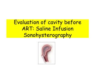
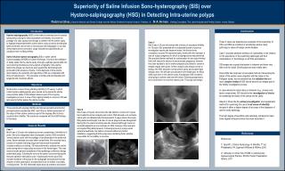
Superiority of Saline Infusion Sono-hysterography (SIS) over Hystero- salpingography (HSG) in Detecting Intra-uterine polyps

E N D
Superiority of Saline Infusion Sono-hysterography (SIS) over Hystero-salpingography (HSG) in Detecting Intra-uterine polyps RubinaIzhar, Karachi Medical and Dental College and the AbbasiShaheed Hospital, Karachi, Pakistan & N A Armar, Visiting Consultant, The Lister Hospital and Fertility Centre, Accra, Ghana. Conclusions Introduction Hystero-salpingography (HSG) is the method commonly used to assess tubal patency during the initial assessment of infertility. It involves the passage of a radio opaque dye through a catheter into the uterine cavity to highlight features/blemishes of the uterine cavity as well as the fallopian tubes and these can be seen on fluoroscopy and radiographs. It can also demonstrate uterine anomalies. Large intrauterine polyps/fibroids can usually be seen as filling defects. Saline infusion hystero-sonography (SIS) or saline uterine hydrosonography (HSGM) is a newer technique. It involves the instillation of sterile saline into the uterine cavity (through a catheter placed within the cervix) during simultaneous trans-vaginal sonography. Intra-uterine lesions like polyps can clearly be seen against the dark background produced by the presence of saline. In the diagnosis of intra-uterine abnormalities, the sensitivity and specificity of SIS are comparable with those of hysteroscopy 1. The procedure is simple and well tolerated and largely devoid of adverse effects. We describe 3 cases of long standing infertility (5-15 years), in which routine hystero-salpingograms were carried out to assess the uterine cavity and the status of the fallopian tubes as part of the ongoing investigation process. In view of the ongoing inability to conceive, SIS was later used to further assess the uterine cavities. Three women with ongoing infertility and whose reproductive tracts had previously been evaluated by HSG had SIS as a further assessment of the status of their uterine cavities as part of the ongoing effort to find a cause for their infertility. The results are compared with the HSG findings in this study. Case 1 Mrs N was a 32 years old nulliparous woman complaining of infertility for 8 years. She was a diagnosed case of polycystic ovaries. HSG revealed a ‘normal uterine cavity’ with free spillage of dye bilaterally in the peritoneal cavity. Semen analysis was also within normal limits. She received many courses of ovulation inducing drugs and had several documented ovulations without conceiving. SIS was performed to evaluate her uterine cavity during which a large polyp was seen in the fundal region. This was removed under general anaesthesia. Histo-pathology confirmed a benign endometrial polyp. Following its removal the patient conceived during the first post-operative stimulation cycle. A subsequent review (post SIS) revealed evidence of the polyp on the radiograph (arrowed) but this was missed on initial examination, and assumed to be an artifact or possibly, overlying bowel. The SIS eliminated doubt about its presence and nature. These 3 cases are presented as examples of the superiority of SIS over HSG as a method of scrutinizing uterine cavity pathology in cases of benign uterine disease. Case 1 provides pertinent evidence of vulnerability to the reliance on the experience of the examining radiologist and their interpretation of the findings on an HSG examination. SIS images are a great deal easier to interpret and leave very little room for doubt. Small polyps are also readily seen. Since HSG has long been an accepted method of assessing the status of the uterine cavity (together with the status of the Fallopian tubes), we recommend that the radiation freeand much simpler method of SIS should become an integral part of the fertility investigation process. In cases where the tubal status is irrelevant (e.g., severe male factor compromise), SIS should be the investigation of choicefor assessing the status of the uterus and its cavity. Indeed, it should be the primary investigationand assessment method for examining the uterus in all cases of infertility because it offers a higher degree of accuracy in the diagnosis of uterine cavity pathology. The high degree of specificity and sensitivity compared to basic trans-vaginal ultrasound alone has been described 2.. Case 3 Mrs S was a 35 year old woman with a history of secondary infertility for 15 years. She presented with a substantial bundle of previous investigation reports and treatment notes. Her first and only conception occurred 14 years previously, shortly after her marriage. It was an ectopic pregnancy for which a salpingectomy was performed. She practiced barrier methods for contraception for two years thinking that it will reduce the chance of repeat ectopic pregnancy. However, they later decided to try for another pregnancy but failed to conceive despite regular intercourse. Semen analysis was always normal on testing. Her HSG showed a normal uterine cavity and a patent right fallopian tube. Her left tube was blocked in the middle part. No filling defect was seen in the uterine cavity. A subsequent SIS revealed a small polyp in anterior wall near the fundus. Following polypectomy, she conceived in her very first natural cycle. The polyp was benign. Objectives Methods Case 2 Mrs Z was a 25 years old woman who had failed to conceive for 5 years. Her husband’s semen analysis was normal. HSG showed a uni-cornuate uterus with one fallopian tube that was patent. A vague linear intra-cavity filling defect was thought to be due to mucus plugs and was disregarded. During SIS, the uterine anomaly was also observed although it was not as readily apparent as one could see on HSG. However, there was also an irregular filling defect present. Following removal of a polyp under general anaesthesia, the patient conceived without any further assistance, suggesting that the polyp was a hindering factor possibly responsible for her inability to conceive. Cases & Results References References: 1/ Speroff L. Clinical Gynecology & Infertility. 7th ed, Philadelphia, PA :Lippincott Williams & Wilkins; 2005 2/ Vathanan V, Armar N A, HSGM in contemporary Gynaecological Practice, RCOG Poster Presentation, Athens, 2011