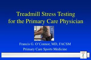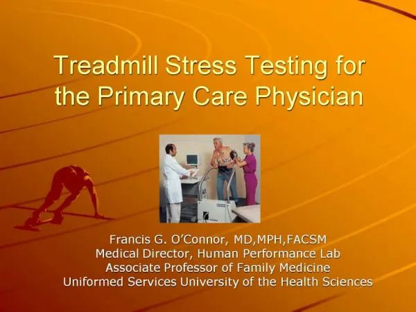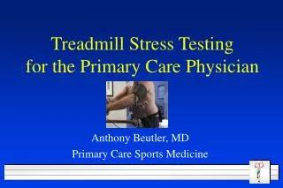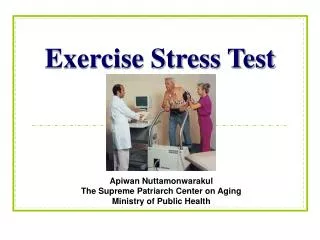
Treadmill Stress Test
Treadmill Stress Test. Majelle L. Gagtan. Performance of the Exercise Stress Test. Definition Indications/Contraindications Running the Exercise Test Protocols. Treadmill Stress Test.

Treadmill Stress Test
E N D
Presentation Transcript
Treadmill Stress Test Majelle L. Gagtan
Performance of the Exercise Stress Test • Definition • Indications/Contraindications • Running the Exercise Test • Protocols
Treadmill Stress Test • Non-invasive procedure providing information about changes in rate, rhythm, conductionn and ventricular repolarization as the heart responds to exertion • Exposes the heart to the stress of exercise thus unmasking s/sx of heart disease, and the ECG may produce characteristic abnormalities
Indications for Exercise Testing • Patients with s/sx suggestive of CAD • Patients with significant risk factors for CAD • To evaluate exercise tolerance in patients with unexplained fatigue and shortness of breath • To evaluate BP response to exercise in patients with borderline hypertension • To look for exercise-induced serious irregular heart beats
Absolute Contraindications to Exercise Setting: • Recent acute MI • Unstable angina • Ventricular tachycardia • Dissecting aortic aneurysm • Acute CHF • Severe aortic stenosis • Active myocarditis • Thrombophlebitis or intracardiac thrombi • Recent pulmonary embolus • Acute infection
Relative Contraindications to Exercise Setting: • Uncontrolled severe hypertension • Moderate aortic stenosis • Severe subaortic stenosis • Supraventricular dysrhythmias • Ventricular aneurysm • Complex ventricular ectopy • Cardiomyopathy • Uncontrolled metabolic disease • Recurrent infectious disease • Complicated pregnancy
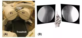
Running the Stress test • HR and BP are recorded at rest • 12L ECG is recorded • Start at a relatively slow “warm up” speed then its speed and inclination are increased every 3 mins. according to a preprogrammed protocol • BP is recorded every minute • Stopped when the patient achieves target HR, or if he develops chest discomfort, dyspnea, dizziness etc., or if the ECG showed significant changes • It may also be stopped if BP rises or falls beyond acceptable limits • Maximum HR = 220 – age of patient
Protocols BRUCE Protocol • multi stage maximal treadmill protocol with 3-min periods to allow achievement of steady state before workload is increased
Protocols Modified BRUCE Protocol • 2 3-min warm-up stages at 1.7mph and 0% grade and 1.7mph and 5% grade • For older individuals or those with exercise capacity is limited by cardiac disease Naughton and Weber protocols • 1 2-min stages with 1 MET increments between stages • More suitable for patients with limited exercise tolerance
Protocols Asymptomatic Cardiac Ishemia Pilot Trial (ACIP) and modified ACIP protocols • For pxs with established CAD • Results in linear increase in HR and VO2 • Modified ACIP – similar aerobic demand; well suited for short or elderly who can’t keep up with a walking speed of 3mph
Positive Negative • ST Depression • → or ↓ ≥ 1mm at 60msec • ↑ ≥ 1.5mm at 80msec • ST Elevation • ≥ 1mm at 60msec • No change • ST depression doesn’t fulfill no.2 • T wave inversion w/o ST segment changes • ST elevatoin in a Q wave lead
When to stop! Dyspnea, fatigue, chest pain Systolic blood pressure drop Technical difficulties ECG--ST changes, arrhythmias Signs of poor perfusion (cyanosis/pallor) Px’s desire to stop Achievement of maximal exercise