Download
1 / 29
320 likes | 788 Views
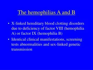
The hemophilias A and B. X-linked hereditary blood clotting disorders due to deficiency of factor VIII (hemophilia A) or factor IX (hemophilia B) Identical clinical manifestations, screening tests abnormalities and sex-linked genetic transmission. Epidemiology. The incidence rate:

E N D
The hemophilias A and B • X-linked hereditary blood clotting disorders due to deficiency of factor VIII (hemophilia A) or factor IX (hemophilia B) • Identical clinical manifestations, screening tests abnormalities and sex-linked genetic transmission
Epidemiology The incidence rate: • Hemophilia A • 1 per 10 000 live male births • Hemophilia B • 1 per 25 000- 30 000 live male births • Found in all ethnic groups, in all parts of the world
Etiology and pathogenesis (1) • Hemophilias result from defects in the factor VIII/IX gene that leads to decreased amount of f. VIII/IX protein, the presence of a functionally abnormal protein, or combination of both
Etiology and pathogenesis (2) • Factor IX activated by the f. VIIa/tissue factor complex or f. XIa forms factor IXa (the active enzyme) • Factor VIII activated by thrombin forms factor VIIIa • Factor IXa activates factor X in the presence of factor VIIIa, phospholipid (activated platelets) and calcium • Factor Xa converts prothrombin to thrombin • In patient with hemophilia, clot formation is delayed because thrombin generation is markedly decreased
Clinical features • Excessive bleeding into various parts of the body • hemarthroses • hematomas • hematuria • hemorrhage into the central nervous system • mucous membrane hemorrhage • pseudotumors (blood cysts) • dental and surgical bleeding
Hemarthroses • Bleeding into joints accounts for about 75% of bleeding episodes in severely affected patients • The joints most frequently involved: knees, elbows, ankles, shoulders , wrists and hips • Repeated hemarthroses results in destruction of articular cartilage, synovial hypertrophy and inflammation • The major complication of repeated bleeding is joint deformity complicated by muscle atrophy and soft tissue contractures
Neurologic complications • Hemorrhage into the central nervous system is the most dangerous event in hemophilic patients • Intracranial bleeding may be spontaneous or follows trauma, which may be trivial • Hemophilic patients with unusual headaches should always be suspected of having intracranial hemorrhage • Hemorrhage into the spinal canal can result in paraplegia • Peripheral nerve compression is a frequent complication of muscle hematomas, particularly in the extremities
Factor VIII/IX activity- definition • 1 unit of factor VIII/IX- equal to the amount in 1ml of pooled fresh normal human plasma • 1 unit of factor VIII/IX/ml is 100% of normal
Laboratory features • Prolonged activated partial thromboplastin time (aPTT) • the aPTT is corrected when hemophilic plasma is mixed with an equal volume of normal plasma • Normal prothrombin time, thrombin-clotting time, bleeding time • A definitive diagnosis of hemophilia A/B should be based on specific assay for factor VIII/IX coagulant activity
Therapy- general principles • Avoidance of aspirin, non-steroid anti-inflammatory drugs, and other agents interfering with platelet aggregation • exception - the pain of hemophilic arthropathy • Addictive narcotic agents should be used with great caution • Avoidance of intramuscular injections
Factor VIII replacement therapy Factor VIII concentrates: • plasma-derived • intermediate purity, high purity and ultrapure concentrates after viral inactivation by pasteurization or by exposure to solvent detergent • produced by recombinant DNA technics
Factor VIII replacement therapy • The site and severity of hemorrhage determine the frequency and dose of factor VIII to be infused • The dose of factor VIII calculation for practical purpose: • 1 unit of factor VIII/kg will raise the circulating f. VIII level about 2% (0.02 U/ml) • after the initial dose of f. VIII further doses are based on a half- life of 8 to 12 h
DDAVP in the treatment of hemophilia A • DDAVP (1,8-desamino-D-arginine vasopressin, desmopressin) causes a transient rise in factor VIII in normal subjects and in patients with mild to moderate hemophilia A • After a dose 0.3 g per kg i.v or s.c. F. VIII level increases two- to threefold above baseline • Repeated administration of DDAVP results in a diminished response (tachyphylaxis)
Factor VIII prophylactic therapy • It should be considered in all severely affected patients • The administration of 20 U factor VIII/kg three times weekly markedly decreases the frequency of hemophilic arthropathy and other long-term effects of hemorrhages episodes
Factor IX replacement therapy Factor IX concentrates: • plasma-derived • intermediate purity- prothrombin complex concentrates • high purity • produced by recombinant DNA technics • viral inactivation: dry heat 80oC, pasteurization, solvent detergent
Factor IX replacement therapy • The dose of factor IX calculation for practical purpose: • 1 unit of factor IX/kg will raise the circulating f. IX level about 1% (0.01 U/ml) • intravascular recovery of factor IX is about 50% (probably f. IX binds to collagen type IV of the vessel wall) • the initial dose of f.IX should be followed by one-half this amount every 12 to 18 h
Antifibrynolytic agents • Fibrynolytic inhibitors (epsilon-aminocapric acid (EACA), tranexamic acid) may be given as adjunctive therapy for bleeding from mucous membranes, particularly for dental procedure • Doses: tranexamic acid (Exacyl) 1g every 6 h EACA 4 g every 6 h
von Willebrand disease • the most common inherited bleeding disorder in humans • results from quantitative or qualitative abnormalities in von Willebrand factor (vWF) • von Willebrand factor is a central component of hemostasis, secreted by endothelial cells, that circulates in plasma in multimers, serving both as a carrier for factor VIII and as an adhesive link between platelets and the injured blood vessel wall
von Willebrand disease- epidemiology • The overall prevalence of von Willebrand disease is 1% of the general population • The prevalence of clinically significant disease is closer to 1: 1000
Classification of von Willebrand disease • Type 1 vWD- the most common variant • autosomal dominant in inheritance • normal vWF in structure and function but decrease in quantity- range 25-50% of normal • Type 2 vWD (2A, 2B, 2M, 2N) • autosomal dominant in inheritance • vWF is abnormal in structure and/or function • Type 3 vWD • autosomal recessive in inheritance • the most severe form characterized by very low or undetectable level of vWF
Clinical symptoms • Mucocutaneous bleeding- the most common symptom • epistaxis • easy bruising and hematomas • menorrhagia • gingival bleeding • gastrointestinal bleeding • spontaneous hemarthroses occur almost exclusively in patients with type 3 vWD
Laboratory features • Screening tests: • bleeding time- normal or prolonged • aPTT- prolonged or normal • PT- normal • The routine tests: • activity of factor VIII- decreased • vWF antigen- decreased • ristocetin cofactor activity assay- decreased agglutination of platelets in the presence of ristocetin • analysis of plasma vWF multimers- critical for subclassification of vWD
Therapy • Desmopressin • a dose 0.3 g per kg i.v or s.c., upper limit 20 g , repeated 3 or 4 times every 24 hours • the best results in type 1 vWD- effective in 80% patients • many patients with type 2 and nearly all ones with type 3 do not respond to DDAVP • vWF replacement therapy • vWF-containing factor VIII concentrates: Humate P, Koate HP
Nonreplacement therapy • Estrogen or oral contraceptives in treating menorrhagia • fibrynolytic inhibitors
The other uncommon inherited deficiencies of coagulation factors • Bleeding tendencies caused by inherited deficiency of factors I, II, V, VII, X, XI and XIII are rare disorders, distributed worldwide • Treatment may be necessary during spontaneous bleeding episodes, during or after surgical procedures • In most deficiency states fresh frozen plasma replacement is used, but specific concentrates of factors I, II, VII, X, XI and XIII are also available