Download
1 / 64
640 likes | 660 Views
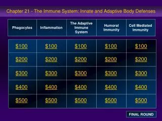
The Immune System: Innate and Adaptive Body Defenses Part A. 21. Immunity: Two Intrinsic Defense Systems. Innate (nonspecific) system responds quickly and consists of: First line of defense – intact skin and mucosae prevent entry of microorganisms

E N D
The Immune System: Innate and Adaptive Body Defenses Part A 21
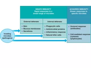
Immunity: Two Intrinsic Defense Systems • Innate (nonspecific) system responds quickly and consists of: • First line of defense – intact skin and mucosae prevent entry of microorganisms • Second line of defense – antimicrobial proteins, phagocytes, and other cells • Inhibit spread of invaders throughout the body • Inflammation is its hallmark and most important mechanism
Immunity: Two Intrinsic Defense Systems • Adaptive (specific) defense system • Third line of defense – mounts attack against particular foreign substances • Takes longer to react than the innate system • Works in conjunction with the innate system
Surface Barriers • Skin, mucous membranes, and their secretions make up the first line of defense
Epithelial Chemical Barriers • Epithelial membranes produce protective chemicals that destroy microorganisms • Skin oils have acidity (pH of 3 to 5)which inhibits bacterial growth • Stomach mucosae secrete concentrated HCl and protein-digesting enzymes • Saliva and lacrimal fluid contain lysozyme
Respiratory Tract Mucosae • Mucus-coated hairs in the nose trap inhaled particles • Mucosa of the upper respiratory tract is ciliated • Cilia sweep dust- and bacteria-laden mucus away from lower respiratory passages
Internal Defenses: Cells and Chemicals • The body uses nonspecific cellular and chemical devices to protect itself • Phagocytes • natural killer (NK) cells • Antimicrobial proteins in blood and tissue fluid • Inflammatory response enlists macrophages, mast cells, WBCs, and chemicals
Phagocytes • Macrophages are the chief phagocytic cells • Free macrophages wander throughout a region in search of cellular debris • Kupffer cells (liver) and microglia (brain) are fixed macrophages There are 3 types of specialized phagocytes. • Neutrophils become phagocytic when encountering infectious material • Eosinophils are weakly phagocytic against parasitic worms • Mast cells bind and ingest a wide range of bacteria
Mechanism of Phagocytosis Figure 21.1a, b
Natural Killer (NK) Cells • Cells that can lyse and kill cancer cells and virus-infected cells • Natural killer cells: • Are a small, distinct group of large granular lymphocytes • React nonspecifically and eliminate cancerous and virus-infected cells • Kill their target cells by releasing perforins and other cytolytic chemicals • Secrete potent chemicals that enhance the inflammatory response
Antimicrobial Proteins • Enhance the innate defenses by: • Attacking microorganisms directly • Hindering microorganisms’ ability to reproduce • The most important antimicrobial proteins are: • Interferon • Complement proteins
Interferon Family • Interferons are a family of related proteins each with slightly different physiological effects • Lymphocytes secrete gamma () interferon, but most other WBCs secrete alpha () interferon • Fibroblasts secrete beta () interferon • Interferons also activate macrophages and mobilize NKs
Interferon (IFN) • Genes that synthesize IFN are activated when a host cell is invaded by a virus • Interferon molecules leave the infected cell and enter neighboring cells • Interferon stimulates the neighboring cells to activate genes for PKR (an antiviral protein) • PKR nonspecifically blocks viral reproduction in the neighboring cell
Interferon (IFN) Figure 21.4
Complement • 20 or so proteins that circulate in the blood in an inactive form • Provides a major mechanism for destroying foreign substances in the body • Amplifies all aspects of the inflammatory response • Kills bacteria and certain other cell types (our cells are immune to complement) • Enhances the effectiveness of both nonspecific and specific defenses
Complement Pathways • Complement can be activated by two pathways: classical and alternative • Classical pathway is linked to the immune system • Depends on the binding of antibodies to invading organisms • Subsequent binding of C1(the first protein) to the antigen-antibody complexes (complement fixation) • Alternative pathway is triggered by interaction among factors B, D, and P, and polysaccharide molecules present on microorganisms
Complement Pathways • Each pathway involves a cascade in which complement proteins are activated in an orderly sequence and where each step catalyzes the next • Both pathways converge on C3, which cleaves into C3a and C3b • C3b initiates formation of a membrane attack complex (MAC) • C3b also causes opsonization (identification of the invader), and C3a causes inflammation
Complement Pathways Figure 21.5
C-reactive Protein (CRP) • CRP is produced by the liver in response to inflammatory molecules • CRP is a clinical marker used to assess for: • The presence of an acute infection • An inflammatory condition and its response to treatment
Fever • Abnormally high body temperature in response to invading microorganisms • The body’s thermostat is reset upwards in response to pyrogens, chemicals secreted by leukocytes and macrophages exposed to bacteria and other foreign substances
Fever • High fevers are dangerous as they can denature enzymes • Moderate fever can be beneficial, as it causes: • The liver and spleen to sequester iron and zinc (needed by microorganisms) • An increase in the metabolic rate, which speeds up tissue repair
Inflammation: Tissue Response to Injury • The inflammatory response is triggered whenever body tissues are injured • Prevents the spread of damaging agents to nearby tissues • Disposes of cell debris and pathogens • Sets the stage for repair processes • The four cardinal signs of acute inflammation are redness, heat, swelling, and pain
Inflammation Response • Begins by Macrophages and other immune cells recognizing an invader. • They do this with TLR’s (Toll Like Receptors), which are receptors on the membrane of the immune cell that can recognize by shape the specific class of microbe.
Chemical Mediators • Once a microbe has been identified a flood of inflammatory chemicals are released into the extracellular fluid • Inflammatory mediators: • Include Cytokines, histamines, kinins, prostaglandins (PGs), and complement. • Cause local small blood vessels to dilate, resulting in hyperemia. • Send out a chemical signal that attracts other white blood cells.
Inflammatory Response: Vascular Permeability • Chemicals liberated by the inflammatory response increase the permeability of local capillaries • Exudate (fluid containing proteins, clotting factors, and antibodies): • Seeps into tissue spaces causing local edema (swelling), which contributes to the sensation of pain
Inflammatory Response: Edema • The surge of protein-rich fluids into tissue spaces (edema): • Helps to dilute harmful substances • Brings in large quantities of oxygen and nutrients needed for repair • Allows entry of clotting proteins, which prevents the spread of bacteria
Inflammatory Response: Phagocytic Mobilization • Occurs in four main phases: • Leukocytosis – neutrophils are released from the bone marrow in response to chemical released by injured cells • Margination – neutrophils cling to the walls of capillaries in the injured area • Diapedesis – neutrophils squeeze through capillary walls and begin phagocytosis • Chemotaxis – inflammatory chemicals attract neutrophils to the injury site
Monocytes • Hours After the neutrophils have reached the injured site monocytes reach the area. • They swell, develop large numbers of lysosomes and become macrophages with insatiable appitites. • If these macrophages can not deal with the situation then a chemical signal will be sent for the adaptive immune system (T and B cells) to come to the rescue.
Platelets and Coagulation • Platelets do not stick to each other or the endothelium lining unless the lining is damaged. • When the endothelium is damaged the collagen fibers are exposed and the platelets adhere to the fibers. • This causes the platelets to undergo a change • They swell • Form spiked processes • Become sticky.
Coagulation • Coagulation is a complicated multistep process that involves over 30 different substances. • However, the process leads to the same end result, an enzyme called thrombin catalyzes the joining of fibrinogen in the plasma into a fibrin mesh around the wound. • This fibrin mesh traps red and white blood cells effectively sealing the damaged vessel.
Clot retraction and repair • With in 30 to 60 minutes of the fibrin mesh being created, platelets induce clot retraction. • Platelets actually contain actin and myosin which allows the platelets to draw both end of the damaged tissue or vessel together. • As this happens healing of the tissue has already begin stimulated by Platelet derived growth factors (PDGF) and Vascular endothelial growth factors (VEGF).
Clot Removal • Once healing has occurred the clot is removed by a fibrin digesting enzyme called plasmin. • Remember while all of this healing is occurring the inflammation process is drawing the necessary immune cells to the are. • The ultimate goal of inflammation is the clear the injured site and aid in tissue repair.
Flowchart of Events in Inflammation Figure 21.2
Adaptive (Specific) Defenses • The adaptive immune system is a functional system that: • Recognizes specific foreign substances • Acts to immobilize, neutralize, or destroy foreign substances • Amplifies inflammatory response and activates complement
Adaptive Immune Defenses • The adaptive immune system is antigen-specific, systemic, and has memory • It has two separate but overlapping arms • Humoral, or antibody-mediated immunity • Cellular, or cell-mediated immunity
Antigens • Substances that can mobilize the immune system and provoke an immune response • The ultimate targets of all immune responses are mostly large, complex molecules not normally found in the body (nonself)
Complete Antigens • Important functional properties: • Immunogenicity – the ability to stimulate proliferation of specific lymphocytes and antibody production • Reactivity – the ability to react with the products of the activated lymphocytes and the antibodies released in response to them • Complete antigens include foreign protein, nucleic acid, some lipids, and large polysaccharides
Haptens (Incomplete Antigens) • Small molecules, such as peptides, nucleotides, and many hormones, that are not immunogenic but are reactive when attached to protein carriers • If they link up with the body’s proteins, the adaptive immune system may recognize them as foreign and mount a harmful attack (allergy) • Haptens are found in poison ivy, dander, some detergents, and cosmetics
Antigenic Determinants • Only certain parts of an entire antigen are immunogenic • Antibodies and activated lymphocytes bind to these antigenic determinants • Most naturally occurring antigens have numerous antigenic determinants that: • Mobilize several different lymphocyte populations • Form different kinds of antibodies against it • Large, chemically simple molecules (e.g., plastics) have little or no immunogenicity
Antigenic Determinants Figure 21.6
Self-Antigens: MHC Proteins • Our cells are dotted with protein molecules (self-antigens) that are not antigenic to us but are strongly antigenic to others • One type of these, MHC proteins, mark a cell as self • The two classes of MHC proteins are: • Class I MHC proteins – found on virtually all body cells • Class II MHC proteins – found on certain cells in the immune response
MHC Proteins • Are coded for by genes of the major histocompatibility complex (MHC) and are unique to an individual • Each MHC molecule has a deep groove that displays a peptide, which is a normal cellular product of protein recycling • In infected cells, MHC proteins bind to fragments of foreign antigens, which play a crucial role in mobilizing the immune system
Cells of the Adaptive Immune System • Two types of lymphocytes • B lymphocytes – oversee humoral immunity • T lymphocytes – non-antibody-producing cells that constitute the cell-mediated arm of immunity • Antigen-presenting cells (APCs): • Do not respond to specific antigens • Play essential auxiliary roles in immunity
Lymphocytes • Immature lymphocytes released from bone marrow are essentially identical • Whether a lymphocyte matures into a B cell or a T cell depends on where in the body it becomes immunocompetent • B cells mature in the bone marrow • T cells mature in the thymus
T Cell Selection in the Thymus Figure 21.7
T Cells • T cells mature in the thymus under negative and positive selection pressures • Negative selection – eliminates T cells that are strongly anti-self • Positive selection – selects T cells with a weak response to self-antigens, which thus become both immunocompetent and self-tolerant
B Cells • B cells become immunocompetent and self-tolerant in bone marrow • Some self-reactive B cells are inactivated (anergy) while others are killed • Other B cells undergo receptor editing in which there is a rearrangement of their receptors
Immunocompetent B or T cells • Display a unique type of receptor that responds to a distinct antigen • Become immunocompetent before they encounter antigens they may later attack • Are exported to secondary lymphoid tissue where encounters with antigens occur • Mature into fully functional antigen-activated cells upon binding with their recognized antigen • It is genes, not antigens, that determine which foreign substances our immune system will recognize and resist
Immunocompetent B or T cells Key: = Site of lymphocyte origin Red bone marrow = Site of development of immunocompetence as B or T cells; primary lymphoid organs = Site of antigen challenge and final differentiation to activated B and T cells Immature lymphocytes Circulation in blood 1 Lymphocytes destined to become T cells migrate to the thymus and develop immunocompetence there. B cells develop immunocompetence in red bone marrow. 1 1 Thymus Bonemarrow 2 After leaving the thymus or bone marrow as naive immunocompetent cells, lymphocytes “seed” the lymph nodes, spleen, and other lymphoid tissues where the antigen challenge occurs. 2 Immunocompetent, but still naive, lymphocyte migrates via blood 2 Lymph nodes, spleen, and other lymphoid tissues Mature (antigen-activated) immunocompetent lymphocytes circulate continuously in the bloodstream and lymph and throughout the lymphoid organs of the body. 3 3 3 Activated immunocompetent B and T cells recirculate in blood and lymph Figure 21.8