General Pathology (PATH 303) Lecture # 10
190 likes | 445 Views
General Pathology (PATH 303) Lecture # 10 Cellular adaptations of growth. Cellular adaptations of growth:.

General Pathology (PATH 303) Lecture # 10
E N D
Presentation Transcript
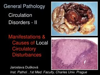
General Pathology (PATH 303) Lecture # 10 • Cellular adaptations of growth
Cellular adaptations of growth: • Cells constantly adapt (adjust) to the changes in their environment. These adaptations maybe physiological or pathological and include atrophy, hypertrophy, hyperplasia and metaplasia. Aplasia: • It is the failure of an organ or tissue to develop. • It occurs in the embryo or foetus during intrauterine development.
Causes: • Hereditary defects in germplasm. • Death of one or a few cells in embryonic stage by viral infection of dam. Gross and Microscopic Appearance: • Organ or tissue is totally absent. • Usually, there is a fatty or fibrous mass. • Aplasia of a vital organ is incompatible with life.
Hypoplasia: • Failure of cells, tissues or organs to attain their mature size. Causes: • Congenital anomalies due to unknown causes. • Intrauterine viral infections during early gestation. • Ingestion of toxic substances and nutritional deficiencies during early pregnancy.
Gross Appearance: • The affected tissues and organs are smaller than normal and fail to attain the adult size. Microscopic Appearance: • The cells in the hypoplastic tissues are smaller in size and number than normal. • There is excessive amount of fibrous connective tissue and fat
Atrophy: • It is a decrease in size of cells (tissues and organs) after they have reached their full development. • There is a loss of cell substance. Causes: According to its causes, atrophy is of several types e.g. Disuse Atrophy: • Due to decreased workload or inactivity – atrophy of muscles occurs when a plaster cast is applied to a broken limb. • Neurogenic or neutrotropic atrophy due to loss of innervation. Atrophy of laryngeal muscles (roaring) occurs in horses due to degeneration of left recurrent nerve.
Endocrine Atrophy: • Due to decrease of hormones (physiologic atrophy) seen in involution of thymus and mammary gland etc. • Nutritional (starvation) atrophy: Caused by starvation or malnutrition. • Angiotrophic or Vascular Atrophy: Caused by partial loss of blood supply. • Pressure Atrophy: Due to mild, continuous pressure on cells and tissues causing obstruction of blood supply or ducts.
Biochemical Mechanism: • Atrophy is caused by an imbalance between synthesis and degradation. There maybe decreased synthesis or increased catabolism or both. • Degradation of protein (proteolysis) plays a key role in atrophy. • The cell has two mechanisms (pathways) for proteolysis: • Lysosomes contain proteases and other enzymes that degrade molecules from old (senescent) cytoplasmic organelles like (mitochondria, ribosomes, endoplasmic reticulum). lysosomal – pathway. • Ubiquitin – Proteosome Pathway: Proteins to be destroyed are conjugated to ubiquitin which is a 76 – amino acid cytosolic peptide. Protein is then destroyed within large cytoplasmic bodies called proteosomes.
Gross Appearance: • The affected organ is decrease in size, soft and flabby and may appear pale and anemic. Microscopic Appearance: • The cells are smaller in size and number. The cytoplasm contains membrane-bound vacuoles which contain fragments of cell components (organelles). These autophagic vacuoles may persist and give a brown discolouration to tissue as in case of lipofuscin granules. Chronic inflammation in many organs is associated with atrophy
Hypertrophy: • An increase in size of a tissue or organ without an increase in number of cells. • Causes: • It is a response to demand for increased workload or from endocrine stimulation. • Physiological hypertrophy is seen in uterus in pregnancy and in muscles in weight lifters. • Adaptive hypertrophy is also seen in cardiac and skeletal muscles in response to increased workload. • Compensatory hypertrophy: It is most commonly seen in the paired organs. If one organ of the pair e.g. kidney is damaged or removed the other organ increases in size to compensate for the loss.
Mechanism: • The size of cells increases due to increase in size and number of myofilaments in the cardiac and skeletal muscles. • A growth factor has been isolated from the hypertrophied muscles.
Gross Appearance: • The tissue or organ is larger and heavier than normal. Microscopically: • The size of cells is increased indicated by a decrease in the number cells in each microscopic field. Significance and Results: • Compensatory hypertrophy increases the function of organ and tissues. If the workload continues to increase decomposition and organ failure will occur.
Hyperplasia: • It is an increase in size of tissue and organ due to an increase in the number of cells. Hyperplasia and hypertrophy are closely associated and may coexist. Causes: • Hyperplasia maybe physiological or pathological: • The main cause of physiological hyperplasia is hormonal stimulation as the proliferation of glandular epithelium of mammary gland in pregnancy. • Compensatory or regenerative hyperplasia is seen when a portion of an organ like liver is removed. The remaining cells undergo mitotic activity due to stimulation by polypeptide growth factor.
Pathological Hyperplasia: • Prolonged, chronic irritation is the main cause of pathological hyperplasia – Irritation maybe caused by mechanical, chemical or thermal agents like rubbing injuries (calluses), chlorinated naphthalene. • Endocrine disturbances e.g. hyperplasia of prostate in old dog which can be treated by castration or treatment with estrogen. • Nutritional disturbances like deficiency of iodine & Vit. A cause goitre in thyroid and hyperkeratinization of epithelium in esophagus in chicken, especially. • Infectious agents e.g. viruses in pox and contagious ecthyma. There is increased sensitivity to normal level of growth factor.
Wound Healing: • Fibroblasts and angioblasts proliferate due to stimulation by the growth factor and facilitate healing and repair of wounds. Significance: • Epithelial hyperplasia usually disappears when the cause is removed, however hyperplasia of FCT persists. • Hyperplasia may give rise to neoplasia (cancer).
Metaplasia: • It is substitution of one fully differentiated adult cell type in a tissue by another adult cell type. Causes: • Repeated and prolonged irritation by lung worm infection or cigarette smoking changes pseudostratified columnar epithelium in trachea and bronchi. • Deficiency of vitamin A causes replacement of cuboidal epithelium in the ducts of esophageal glands with stratified squamous epithelium. • Hormonal Disturbances: Mammary gland neoplasms are caused by hormonal disturbances in dogs and squamous epithelium, cartilage, bone and muscle tissues are seen in such tumours (mixed mammary gland tumours).
Gross and Microscopic Appearance: • One adult type of tissue is replaced by another well-differentiated tissue and gross and microscopic appearance are seen accordingly e.g. cartilage and bone maybe present in mammary gland. Significance: • Metaplasia is an adaptive response in which cells sensitive to a particular stress are replaced by more resistant cell type. • Metaplastic tissue returns to normal if the cause is removed, however cartilage and bone remain permanently. • Metaplasia may lead to neoplasia.