Unit H
Unit H. Diagnostic Skills. Objectives. 2H08.01 Measure and record vital signs 2H08.02 Apply medical assisting and laboratory skills. Measure and record vital signs. Temperature Converting: To convert Fahrenheit (F) to Celsius (C) : (F – 32) 5/9 To convert Celsius To Fahrenheit :

Unit H
E N D
Presentation Transcript
Unit H Diagnostic Skills
Objectives • 2H08.01 Measure and record vital signs • 2H08.02 Apply medical assisting and laboratory skills
Measure and record vital signs Temperature Converting: To convert Fahrenheit (F) to Celsius (C) : (F – 32) 5/9 To convert Celsius To Fahrenheit : (C X 9/5) + 32
Factors that influence Temperature Factors that rise body temperature: • Illness • Infection • Exercise • Excitement • High temps. In the environment
Factors that Lower the body temperature: • Starvation or fasting • Sleep • Decreased muscle activity • Exposure to cold in the environment • Certain diseases
Solve The Problem • A nurse goes in to check a resident’s oral temperature. She notices that the resident is currently eating their dinner. What should the nurse do?
Solve The Problem • Answer: • You should come back 15 mins. Later after the resident is done eating.
Abnormal Conditions Hypothermia: Below 95 degrees F. Caused by prolonged exposure to cold. Death when temp. is below 93 degrees F. Fever: Elevated temperature, above 101 F Hyperthermia: Temperature above 104 degrees . Caused by prolonged exposure to hot temps., brain damage, or serious infection. Temps above 106 can lead to convulsions and death.
Taking Temperatures • Electronic: can be used for oral, rectal, axillary, or groin. • Tympanic: Placed in auditory canal, taker pushes the scan button. Known as an aurel temperature.
Pulse • Pulse sites: • Radial Artery • Brachial Artery • Temporal Artery • Carotid Artery • Femoral Artery • Popliteal Artery • Doralis pedis Artery
Pulse Terminology • Bradycardia: under 60 beats per minute • Tachycardia: over 100 beats per minute • Rhythm: Regularity of the pulse • Volume: Strength or intensity • Apical Pulse: taken with a stethoscope at the apex of the heart.
Respirations • Character- depth and quality of respirations: • Deep • Shallow • Labored • Difficult • Stertorous • Moist
Abnormal Respirations • Dyspnea- difficult or labored breathing • Apnea- absence of respirations • Cheyne-Stokes: periods of dyspnea followed by periods of apnea. • Rales- bubbling or noisy sounds caused by fluids or mucus in the air passage
Measuring Respirations • Normal rate = 14 – 18 min • Leave your hand on the pulse while counting respirations and be sure the patient doesn’t know you are counting the respirations.
Blood Pressure • Systolic: Pressure on the walls of arteries when the heart is contracting. Normal range- 100 to 140 mm Hg~Top #120/80 • Diastolic: Constant pressure when heart is at rest. Normal range- 60 to 90 mm Hg • Sphygmomanometers- usually aneriod or mercury~ Bottom #100/60
Factors that influence blood pressure • Rise blood pressure: • Excitement, anxiety, nervous tension • Stimulant drugs • Exercise and eating • Lower Blood Pressure: • Rest or sleep • Depressant drugs • Shock • Excessive loss of blood
Apply medical assisting and laboratory skills Principles of height and weight measurements: • Use the same scale everyday, make sure scale is balanced, weigh at the same time each day, and make sure patient is wearing the same amount of clothing each day.
Measuring/Recording Height and weight • Used to determine if patient is underweight or overweight. • Height/Weight chart is used as averages • + or – 20% is considered normal • Patients with edema are weighed daily. • Clinical scales contain a balance beam and measuring rod.
Infant Height and Weight • Keep one hand slightly over but not touching the infant when weighing it. • To measure the infant’s height, mark the exam table at the top of the head and at the bottom of the stretched out heel, then measure mark to mark.
Positioning Patient • Medical exam table • Surgical table • Bed • After use clean table, observe safety factors, and protect the patients privacy. • Be sure to observe patient for signs of distress.
Positioning Patient Horizontal Recumbent/Spine Assist patient to lie on back with arms at side. Use small pillow for head. Patients with lower abdominal pain are placed in this position.
Prone Turn patient toward self. Position patient on abdomen with arms flexed by head. Position head to side on small pillow. This is used during spinal exams.
Positioning Patient Sims / Left Lateral: Turn patient on left side. Position left arm behind back, right arm bent at elbow in front of body. Turn head to side on small pillow. Flexed left leg slightly, right leg to abdomen.
Fowlers Position patient on back. Elevate head & back 25 degrees for low-Fowlers. 45 degrees for semi-Fowlers and 90 degrees for high-Fowlers. Flex knees slightly and supports with pillow. This position may help with patients who are having difficulty breathing.
Lithotomy Place patient on back, arms at side, buttocks at end of table. Place pillow under head. Flex and separate knees and place feet in stirrups. • Used during gynecological exams
Trendelenburg: Position patient on back, incline table with head lower than body.
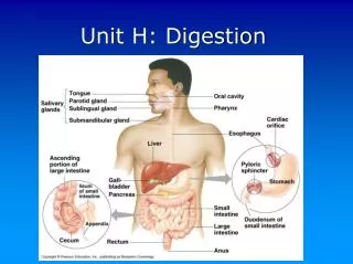
Medical Laboratory Skills Testing Urine- • Urinalysis: usually consists of physical, chemical and microscopic tests. • Physical: color, odor, transparency and specific gravity. • Chemical: to check pH, protein, glucose, ketone, bilirubin, urbilinogen, and blood. • Microscopic: to look for casts, cells, crystals, and amorphous deposits.
Urinalysis • Be sure the specimen is fresh • Cells are normally found during microscopic exams. • Reagent strips test for cellular properties • Physical Properties Measured: • Odor • Transparency • Specific gravity • Color Reagent Strips