Download
1 / 45
620 likes | 1.33k Views
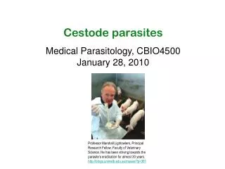
Cestode parasites Medical Parasitology, CBIO4500 January 28, 2010. Professor Marshall Lightowlers, Principal Research Fellow, Faculty of Veterinary Science. He has been striving towards the parasite’s eradication for almost 30 years. http://blogs.unimelb.edu.au/musse/?p=381. Turbellarians.

E N D
Cestode parasites Medical Parasitology, CBIO4500 January 28, 2010 Professor Marshall Lightowlers, Principal Research Fellow, Faculty of Veterinary Science. He has been striving towards the parasite’s eradication for almost 30 years. http://blogs.unimelb.edu.au/musse/?p=381
Turbellarians Free-living worms Monogenea Monogenetic flukes Trematodes Digenetic flukes Cestodes Tapeworms Helminths Phylum Platyhelminths Phylum Nematoda
Review Schistosomes • They live in the bloodstream • They live for a long time • The host can become resistant to infection • The parasites do not replicate in their definitive host • Pathology is not due to host response against the worms but rather to eggs entrapped in tissues • Infect 200 millions of the world’s population in Africa, Asia and S. America. Population at risk: 600 millions • Disease: schistosomiasis or Bilharzia • They need a snail IH for transmission
Tapeworms: the Cestodes • They are flat in cross section • Hermaprhoditic • Live in the intestines with feces • Life cycles are complex and can include multiple intermediate hosts • No mouth, no digestive system • They have suckers and teeth that grasp the host. • Reproductive structures • Behind a short neck are repeated parts of the worm, each containing reproductive structures with eggs and sperm, which can be released with the host's feces. • The pieces give the worm a ribbon-like structure, beneficial for absorbing nutrients from the intestine. http://www.ndpteachers.org/perit/biology_image_gallery1.htm
Order pseudophyllidea: have scolex with bothria; the sperm whale tapeworm, H. physesteris, can be > 30 m long; the genital pore and uterine pore are located on the mid-ventral surface, and the ovary is bilobed ("dumbbell-shaped"); each segment has 4-14 complete sets of genitalia, can be up to 45,000 segments in a worm • Diphyllobothrium sp. • Order cyclophyllidea: most important group of tapeworms of humans and domestic animals, multiple proglottid, scolex ("head") with four suckers. Proglottids have genital openings on one side (except the familyDilepididae, which has them on both sides), and a compact yolk gland or vitellarium posterior to the ovary; can be small (a few mm's) or large (up to 10 m); mammals serve as intermediate hosts • Family Taeniidae:Taenia saginata, Taenia solium, taenia multiceps; Echinococcus granulosus, E. multilocularis • Family Hymenolepididae: Hymenolepis diminuta, Hymenolepis nana, CESTODES Diphyllobotrium latum
HOSTS • The tapeworm life cycle involves a definitive host and one or more intermediate hosts (IH). Exception: Hymenolepis nana only uses one host and D. latum has 2 IHs • INTERMEDIATE: ingests the eggs which develop into larval forms and later into encysted forms in extraintestinal tissues. Each parasite species has specialized larval forms. • DEFINITIVE: Harbors the mature forms of the parasite. Carnivorous or omnivorous mammal that acquires infection by consuming larval cysts in the uncooked tissues of an IH
scolex neck strobila Tapeworms • The body plan of adult cestodes includes a scolex (looks like the “head”), a neck and strobila that can extend for only a few proglottids or thousands • The strobila is not truly metameric though as several organs like the excretory system extend through the entire worm • Proglottid: each individual segment • Most worms are very long: occupying the entire length of small intestine
Morphology of tapeworms: The scolex Scolex of Diphyllobothrium latum. The scolex is the part of the worm that anchors it to the intestinal epithelium and prevents that the worm is passed with the digested food Scolex of Taenia Solium. http://www.denniskunkel.com/product_info.php?products_id=813 The scolex structure varies between species of tapeworms. a. Diphyllobothrium latum has a scolex with elongated, slit-like attachment organs (bothria) b. Taenia saginata has four muscular SUCKERS. c. Taenia solium has similar muscular SUCKERS and a ROSTELLUM with rows of chitinous hooks. Scolex of Taenia Saginata
Strobila • In most cestodes the scolex is tiny when compared to the strobila which makes up most of the actual “worm” • The strobila consists of a linear series of proglottids • Tape worms are hermaphrodites and each proglottid carries a set of female and male reproductive organs • These segments are released and are eliminated with the feces of the host. *(Magnifications are based on a 35mm slide image of 24mm in the narrow dimension.) http://www.denniskunkel.com/product_info.php?products_id=813
Strobila • Strobilation:asexual process of forming segments • New proglottids are continuously formed in the neck just below the scolex (A) • Along the length of the worm the proglottids increase in size and maturity, developing from premature (B) to mature (C, carrying fully functional and active sexual organs), to the “gravid” stage (D) in which essentially the entire proglottid is filled with the uterus and eggs
The tegument • Cestodes do not have a mouth or any form of intestine • The entire uptake of nutrients occurs through multinucleate syncytial tegument. • In reflection of this important role in uptake the absortive surface is highly enlarged by small microvilli or microtriches • Microfilaments (actin polymers) are the molecular backbone of microtriches
With very few exceptions vertebrates are the final host harboring the adult tape worms • Many invertebrates and vertebrates are parasitized as intermediate hosts • The embryonate egg contains the oncosphere a larva that will penetrate the intestinal wall after eggs are swallowed by intermediate host • The oncospheres of eucestoda have three pairs of hooks which makes it easy to identify them Diagram of oncosphere of Hymenolepis diminuta, dorsal view The egg of the pseudophyllidean tapeworm (left) has a thin shell wall and an operculum, which on hatching opens to release the free swimming larvae. In contrast, the egg of the cyclophyllideans tapeworms (right) has a very thick, resistant egg shell, with no operculum. The eggs of T. saginata and solium are similar. CDC Eggs larval stages (IH) adult forms (DH) Eggs Developmental stages: the egg
Developmental stages • The zygote develops into an oncosphere • In some species with aquatic hosts the inner envelope develops into a ciliated epithelium. • These motile coracidia have to be taken up by an intermediate host within a short time • Once inside the gut of the host the oncosphere sheds the ciliated layer and invades and differentiates into a procercoid
(D. latum) (D. latum) (T. solium) (Hymenolepis sp.) (E. granulosus) Developmental stages (metacestodes) • In pseudophyllidean cestodes the larvae look fairly similar to the adult. The first host is infected by a procercoid which still carries the larval hooks • In the second host a plerocercoid forms (there is no asexual amplification) • The cyclophyllidean larvae are more complex and come in a quite a variety • The medically important larvae are cysticercoid, cysticercus and hydatid (some of these larvae provided amplification)
Developmental stages • Most cyclophyllidean cestode larvae (or metacestodes) are some form of a liquid filled bladder with an invaginated scolex (and this theme is the varied in many ways) • In most cases the adult is very well adopted to the host and causes no damage, it is the larvae that are dangerous pathogens The cyst was defined as a hydatid cyst after the pathological diagnosis. Having been reevaluated as a primary focus, liver profile and abdominal ultrasonography findings were normal; and serological tests results were negative. Hydatid cyst in inguinal region was accepted as a primary cyst. Gulten Kiyak, Mehmet Ozer, Recep Aktimur & Ahmet Kusdemir: Primary Hydatid Disease of the Soft Tissue: The Internet Journal of Surgery. 2006; Volume 8, Number 2.
Eggs larval stages (IH) adult forms (DH) Eggs Development in the Definitive host • In the small intestine of the DH, the juvenile worm excyst, evaginate, or both. • Digestive enzymes from the host ‘s gut may play a role in the release of the organisms from their cyst. Temperature is also important. • Development of the strobila is influenced by size of the juvenile, worm species, size and diet of the host, presence of other worms, and immune and/or inflammatory state of the host • Carbohydrates in the diet are important for the growth of the worm. Polysaccharides able to release glucose are ideal. Glucose or a disaccharid containing glucose, such as sucrose are not as good. • As a worm approaches maximal size, growth rate decreases. • T. saginata may live in a human for more than 30 years.
Order pseudophyllidea: have scolex with bothria; the sperm whale tapeworm, H. physesteris, can be > 30 m long; the genital pore and uterine pore are located on the mid-ventral surface, and the ovary is bilobed ("dumbbell-shaped"); each segment has 4-14 complete sets of genitalia, can be up to 45,000 segments in a worm • Diphyllobothrium sp. • Order cyclophyllidea: most important group of tapeworms of humans and domestic animals, multiple proglottid, scolex ("head") with four suckers. Proglottids have a yolk gland or vitellarium posterior to the ovary; can be small (a few mm's) or large (up to 10 m); mammals serve as intermediate hosts • Family Taeniidae:Taenia saginata, Taenia solium, taenia multiceps; Echinococcus granulosus, E. multilocularis • Family Hymenolepididae: Hymenolepis diminuta, Hymenolepis nana, CESTODES Diphyllobotrium latum
Diphyllobotrium latum - the fish tape worm-Life cycle • Common in fish eating carnivores with little host specificity. Salmon, trout, perch, white fish, eel, pike, etc. • Adults get quite long (10 m) and shed up to a million eggs per day • Eggs must reach water for embryonation • After several days a coracidium hatches through the operculum and is eaten by a copepod http://www.dpd.cdc.gov/DPDx/HTML/Diphyllobothriasis.htm
Diphyllobotrium latum - the fish tape worm • The coracidium is eaten by the copepod (First Intermediate Host) • It looses its ciliated coat and once through the intestine and into the hemocoel, it develops into the procercoid in 3 weeks ( 500 m) with a cercomer at the posterior end • The copepod is weakened by the parasite and less motile • The procercoid can not develop any further until is eaten by a fish. • The larvae penetrate the small fish gut (Second Intermediate Host) and migrates in the muscle • Here it grows and matures into the plerocercoid
Diphyllobotrium latum - the fish tape worm • Mature plerocercoids can be easily seen as white masses in uncooked fish • If host fish is eaten by other fish plerocercoids will migrate into muscle of new fish host (paratenic host) • The plerocercoid develop into immature and then into mature adult tapeworms in the small intestine. • About 9 million people infected (was wide spread in northern Europe and Japan, more cases in the US due to sushi, sashimi and ceviche) • Infection occurs through raw fish dishes or handling (tasting) of fish dishes before cooking • Infection rate can be locally quite high, especially when untreated sewer is led into lakes (there are also reservoirs in many carnivores)
Diphyllobotrium latum - the fish tape worm • Definitive host:fish-eating carnivores:dogs,bears, humans, etc. • Infection of humans cause no or little symptoms (abdominal discomfort, nausea diarrhea are rare) • The parasite takes up large amounts of Vitamin B12 • In patients with genetic deficiencies in Vit B12 uptake the parasite competes effectively for the entire vitamin leading to severe pernicious anemia • Geographic distribution: Northern Europe, Chile, Japan, Korea North America Diphyllobothrium latum (broad fish tapeworm) http://animaldiversity.ummz.umich.edu/site/resources/Grzimek_inverts/Cestoda/Diphyllobothrium_latum.jpg/view. html
Diphyllobotrium latum - the fish tape worm • Diagnosis through detection of characteristic eggs in feces • These eggs are oval or ellipsoidal an operculum (arrows) at one end that can be inconspicuous. At the opposite end there is a small knob that can be barely discernible • The eggs are passed in the stool unembryonated. Size range: 58 to 76 µm by 40 to 51 µm. • Treatment with praziquantel • Proglottids of Diphyllobothrium latum. These proglottids tend to be passed in strands of variable length in the stool. The proglottids tend to be broader than long. Image contributed by Georgia Division of Public Health.
TAENIA SAGINATA • Beef tapeworm • Ranges in length from 6-30 ft • Geographic distribution: cosmopolitan. Most common where poor sanitation and no inspection of meat combine • Africa and South America • Transmission: Ingestion of larval form in undercooked beef • In N. America 1 in 100 is infected. In third-world nations could be up to 10% • No symptoms or some abdominal discomfort • Diagnosis: finding eggs or proglottids in feces Taenia saginata adult worm. (A) Adult T. saginata in the ileum of a 25-year-old patient. Reflux of barium into the terminal ileum during a barium enema examination revealed a markedly elongated ribbon-like radiolucent shadow representing the adult tapeworm. (B) Adult T. saginata recovered intact following its passage after a vermifuge was administered. Note the extraordinary length of this worm, which may at times reach 20-30 feet.
Taenia life cycle • Humans are the only DHs • The eggs can survive for days to months in the environment • The adult worm attaches by their scolex to the human small intestine. • The adults produce proglottids passed with stool • The eggs are released after the proglottids are passed with the feces
TAENIA SOLIUM The armed scolex of T. solium (note hooks on top of scolex). CDC • T. solium has a scolex (A) with four suckers and a double crown of hooks, a narrow neck, and a large strobila (2-4 m) (B) consisting of several hundred proglottids. • About 2 months after ingestion, proglottids begin to detach from the distal end and are excreted in the feces. • Each segment contains 50-60,000 fertile eggs. Taenia pisiforme, Scolex. Hakenkranz ähnlich dem der T. solium (Mit freundlicher Genehmigung Roche AG): http://www.infektionsnetz.at/test/bilder/mikroskop/taenia_pisiforme_r.jpg The Lancet (2003) 361: 547
TAENIA SOLIUM • Endemic in less developed countries where pigs are raised as food source. Latin America, most of Asia, sub-saharan Africa, and parts of Oceania. • Infection with the adult forms of the parasite produces similar symptoms to infection with T. saginata. Cysticerci: (A) as seen in infected pork; (B) excised into a Petri dish. The white dot in each cyst corresponds to the scolex. The Lancet (2003) 361: 547. The Lancet (2003) 361: 547
Human cysticercosisWhen humans plays the role of the Intermediate Host • Larval stages develop in the human host • Humans acquire cysticercosis through faecal-oral contamination with T. soliumeggs • The oncosphere in the eggs is released by the action of gastric acid and intestinal fluids • Cross the gut wall and enter the bloodstream • They are carried to the muscles and other tissues • They encyst as cysticerci at small terminal vessels (1 cm) (A) and (B) • Neurocysticercosis and ophtalmic cysticercosis MRI of multiple cysts. Image courtesy of the Centers for Disease Control and Prevention. Racemose Cysticercosis-MRI
Neurocysticercosis • The parasite infects the CNS • Epileptic seizures (58-80% when parenchymal brain cysts). • Intracranial hypertension, hydrocephalus, or both. This syndrome is related to the location of parasites in the cerebral ventricles or vasal cisterns. • Occasionally a cyst may grow larger (giant cyst) • Racemose form: high mortality. Large translucent vesicle lobulated without scolex which develops in the basis of the brain or in the ventricles. Sometimes several small vesicles surround a pedicle like a bunch of grapes. • Geographical variation in clinical manifestations From: NEJM (2001) 345:879 Neuroimaging: MRI of viable (A) and degenerating (B) cysts and CT of calcified cysticerci. The Lancet (2003) 361: 547
Cysticercosis diagnosis • Serologic diagnosis: • Antibody assays for cysticercosis: 8 kDa antigens, GP50, FAST-ELISA with the 8 kDA antigen • Antigen-detection assays: circulating antigens means live parasites. Ongoing viable infection. Monoclonal antibodies seem to detect AGs in CSF. • Antibody assays for taeniasis: TSE33 and TSE38 were recognized by a panel of taeniasis but not cysticercocis, patient serum samples. • Neuroimaging diagnosis: CT and MRI provide objective evidence on number and location of cysticerci. Also their viability and the severity of the host inflammatory reaction. MRI showing parenchymal (A) and extraparenchymal (basal ccs) (B) viable NCC. MRI showing calcified cyst with surrounding edema
Cysticercosis treatment • Treatment should be individualized based on cyst location, level of inflammation and clinical presentation • Therapy should include analgesics, antiepileptic drugs, cysticidal drugs, surgical resection of lesions and placement of ventricular shunts • Parenchymal cysticercosis with viable cysts: Albendazole 15 (mg/kg/day) with dexamethasone (0.1 mg/kg/day). Praziquantel. • Subarachnoid ccs: antiparasitic therapy • No reason to use antiparasitic drugs to treat dead calcified cysts. Symptomatic therapy. • Surgical therapy: ventricular shunting to resolve hydrocephalus. Also excision of giant cysts or intraventricular cysts Albendazole
Transmission • It is not possible to acquire NCC by eating pork! • Ingestion of infected pork only causes adult tapeworm infestation: taeniasis. WHY? • Infected pork contains only the larval cysts that develop into adult worms in the human intestine • What is that transmits CCS? • The eggs • Most common source of infective eggs? • A symptom-free tapeworm carrier in the household
Echinococcosis • Echinococcus multilocularis: alveolar echinococcosis.Invasive solid lesions of firm consistency, full of connective tissue and a jelly-like material. • Echinococcus granulosus: cystic echinococcosis.Produces cystic lesions
Echinococcus granulosus -the dog tape worm • Adult E. granulosus adult worms live in the intestine of dogs • They produce eggs which are shed with the feces • Eggs are infective to herbivores (and humans)
Echinococcus granulosus • The oncosphere penetrate intestine of intermediate host and develops into a hydatid • Hydatides are spherical fluid-filled cysts surrounded by a granuloma formed by the host
The Hydatid Cyst • The cyst is lined by a multilayer parasite tissue with the innermost layer being the germinal layer • This layer is a undifferentiated “stem cell” layer that can spawn the formation of “brood capsules” which are themselves lined by GL • The daughter cysts (the encircled body) "bud" into the center of the fluid-filled cyst. • This is a very small portion of the cyst which may become quite large. • Each of the smaller bodies will develop into diminutive tapeworms should this be eaten by a definitive or final host such as a canine.
The Hydatid Cyst • Thousands of protoscolices can fill the hydatid (hydatide sand) • Protoscolices are the infective stage for dogs • Hydatides usually grow slowly but steadily (1-5 cm per year) • They are usually well tolerated until their size becomes a problem or they rupture • Cyst rupture or leakage can result in allergic reactions and metastasis
Echinococcosis: Cystic hydatid disease • Hydatides can be found in several organs but are most frequent in the liver Hydatid cyst in human: http://cal.vet.upenn.edu/dxendopar/parasitepages/cestodes/e_granulosus.html
Echinococcosis: Cystic hydatid disease • Liver cysts cause liver swelling, right epigastric pain, nausea, vomiting • Obstruction of bile ducts and blood vessels can cause cholangitis, jaundice, cirrhosis and portal hypertension This upper abdominal CT scan shows multiple cysts in the liver, caused by echinococcus. Note the large circular cyst (seen on the left side of the screen) and multiple smaller cysts throughout the liver. http://www.drkoop.com/ency/93/ImagePages/1177.html
Echinococcosis: Cystic hydatid disease • Lung cyst are often well tolerated but obstruction and or rupture can cause chestpain, cough and dyspnea • The first symptoms of brain cyst is often focal epilepsy • Diagnosis is by serology, radiology, CT scans and sonograms. • Treatment is surgical. Prognosis depends on size and location of hydatide (mortality is around 5-10%) • Hydatide is often injected with sterilizing fluids to avoid “metastases” • Benefit of chemotherapy is inconsistent
Echinococcosis control • Sylvatic and domestic strains. • Strains adapted to dogs & sheep are more aggressive upon human infection • Ecchinococcosis can be locally quite important • Control of feral dogs, limit access of dogs to sheep offal, treat pet dogs regularly • Effective control programs in many countries including New Zealand, Tasmania, Cyprus and Iceland
Echinococcus multilocularis - the fox tape worm • Sylvatic zoonosis in Europe and northern America • Fox is final host, life cycle similar to E. granulosus • Humans get infected by eating contaminated berries and mushrooms collected in forests populated by foxes
Hymenolepis nana -the dwarf tape worm • Hymenolepis nana occurs relatively frequently world wide and is usually an infection of children • An intermediate host is not required and autoinfections occur frequently • Cysicercoids develop in the lymphatics of villi • Alternatively infection through cysticercoids in insects that contaminate grains or cereal
Hymenolepis nana -the dwarf tape worm • Usually asymptomatic (very high burden can lead to unspecific gastrointestinal symptoms • Infections are cleared with adolescence • Diagnosis by demonstration of characteristic eggs • (accidental infections with H. diminuta the rat tape worm)
http://animal.discovery.com/videos/monsters-inside-me-meter-long-tapeworm.htmlhttp://animal.discovery.com/videos/monsters-inside-me-meter-long-tapeworm.html