Download
1 / 44
480 likes | 587 Views
Cell and tissue culture Model cells in medicine - Stem cells – Tissue engineering. C ell culture = propagation of cells outside the organism = in vitro (Cells in the organism = in vivo ). ADVANTAGES : cellular environment can be manipulated well defined cell type

E N D
Cell and tissue cultureModel cells in medicine - Stem cells –Tissue engineering
Cell culture = propagation of cells outside the organism = in vitro (Cells in the organism = in vivo) • ADVANTAGES: • cellular environment can be manipulated • well definedcell type • large quantities of cells can be obtained • many cellular functions can be investigated • animal experiments can be replaced • living cells can be tested • human cells can be tested • DISADVANTAGES: • does not mimic the complexity of the human body • spontanous evolution
TYPES OF CULTURED CELLS: • primary culture • cell line • finite • continuous • cell strain
1 day spinal cord explant + lymphatic fluid axons grow in culture! Primary culture • Cells derived directly from tissues • A culture from the time of isolation • until its first subculture Developed at first in 1907:
cultured neuron extending processes
Cell line • Derived from primary culture via subcultivation (passage) dissociate cells medium plate cells tissue explant cells dish • Grow and divide while adhering (monolayer) to plastic dishes • Require growth factor for growth • Passaging: after physical or enzymatic dissociation of monolayer cultures the cells are reseeding in lower density
Cell lineFinite cell line • Finite lifespan • A cell line that survives for a fixed number of population doublings, usually ~ 40–60 (Hayflick’s limit), before senescing and ceasing proliferation.
Cell lineContinuous cell line Features: • Immortal (infinite lifespan, over 100 population doublings) • Genetic abnormalities accumulate (transformed cells) • Unlimited growth • Loss of contact inhibition • Loss of adherence dependency (suspension) • Injected in mice may form tumor Transformation can be spontanous or induced by viruses, carcinogens or radiation • May derive: • from finitecell line(in vitro transformation) – e.g. NIH3T3, CHO • fromtumors (in vivo transformation) – e.g. HeLa
Cell strain • A subpopulation of cell lines that have been purified by physical separation, selection or cloning • Which hasspecific defined characteristicse.g. marker chromosome, virus resistance, antigen expression
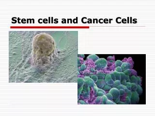
normal cells SEM transformed cells
Requirements of cell cultures • Physical parameters • Temperature • O2/CO2. • humidity • osmolarity • Biological parameters • ASEPTIC conditions • Cell density • Co-culture • Metabolites • Feeder cell layer • Chemical parameters • defined medium • serum (growth factors, hormones) • coat on culture surface (collagen, gelatine) • pH
Laminar box (laminar flow cabinet) is a carefully enclosed bench designed to prevent contamination of biological samples or any particle sensitive materials.
CO2 Incubator The incubator maintains optimal temperature, humidity and other conditions such as the carbon dioxide (CO2) and oxygen content of the atmosphere inside.
Cell cultures can be obtained e.g. from cell banks where these cultures are kept frozen (-196 °C in liquid nitrogen)
Inverted Microscope Light source Objectives in routine laboratories for live cell inspection
R1 ES cells on fibroblast feeder layer D3 ES (embryonic stem) cells on fibroblast feeder layer
myeloma cells HeLa 53 MONOLAYER CELL CULTURE Adherent cells, they need surface for proliferation. These are normal cells except hematopoetic cells. Tumor cells may grow in monolayer, too. SUSPENSION CELL CULTURE Non-adherent, floating, generally hematopoetic or transformed, tumor cells, they do not need special surface for proliferation.
Cell count in function of time Confluent culture
Cell count in function of time 3. Stationary phase /plateau without changingmedium 2. log phase Cell count subculturing 1. lag phase time
ENVIRONMENTAL INTERACTIONS • Infections (viruses, bacteria, • parasites) • Toxicology • Immunology • Carcinogenesis • Biotransformation of xenobiotics • GENETICS • Transformation • Cell fusion • Cell cycle • BIOCHEMISTRY • DNA transcription • RNA metabolism • Protein synthesis • Intermediate metabolism • BIOTECHNOLOGY/ • TISSUE ENGINEERING • Cytokines/growth • factors, hormones, • antibody production • Arteficial tissues • CELL BIOLOGY • Cell-cell and cell-matrix interactions • Gene expression • Cell proliferation • Differentiation • Cell migration, invasion
Slide 9. HeLa monolayer (human cervical cancer)(MGG) cytoplasm nucleus No contact inhibition Cell morphology changes with cell density!! 100% aneuploid.
Research - Infection model Adhesion of different Lactobacilli strains to HeLa cells
Research - Infectionmodel 1 hour 5 hours 22 hours MDCK (Madine Darby canine kidney) cells cultured on microbeads to demonstrate the time course of influenza infection by immunocytochemistry
Diagnosis - Amniocentesis genetic study of the embryo
Therapy - Ex vivo gene therapy In vivo gene therapy
Organ on a chip technique • Microfluidic chamber lined by living human cells • Mimics the interconnectedness of organs within humans • Fields of application: • - disease modelling; • - drug development; • - personalized medicine https://wyss.harvard.edu/technology/human-organs-on-chips/ http://www.thelatestnews.com/organ-chip-breakthrough-replace-lab-animals/
Pluripotent Pluripotent Unipotent
Artificial human tissues and organs • Blood vessels - aorta • Liver • Bone • Cartilage • Skin • Retina Requirements of tissue engineering
Human epidermal epithelial cell cultures with different density and different duration in culture. Cultured epidermal autograft (e.g. Epicel R)
Therapeutic use of human epidermal epithelial cell culture. This photo was taken 5 years after the transplantation.
TISSUE ENGINEERING How to construct an artificial tissue Gene delivery Biopsy Protein secretion Osteogenetic factor Addition of cells Scaffold Implantation
A tissue-friendly scaffold covered with self-cells is not immunogenic.
The immortal life of Henrietta Lacks • 1951 Tissueweretakenwithoutherknowledge • 1952 HeLacellswereusedtodeveloppoliovaccine • 1955Isolation of a singlecellforcloning • 1960 HeLawenttospacebeforeanyastronaut • 1984 HeLawasusedtoprovethatHPV infection causescancer • 1986 Mechanism of HIV infectionwerestudied • 1989 TelomeraseweredescribedinHeLa • 1993 Tuberculosiswasstudied • 2013 Wholegenomedata of HeLawerepublished born Loretta Pleasant in 1920 George Otto Gey
History of human embryonicstemcellresearch 1998 – James Thomson isolated cells from the inner cell mass of the early embryo and developed the first human embryonic stem cell lines Derivation of the H9 cell line. (A) Inner cell mass–derived cells attached to mouse embryonic fibroblast feeder layer after 8 days of culture, 24 hours before first dissociation. Scale bar, 100 μm. (B) H9 colony. Scale bar, 100 μm. (C) H9 cells. Scale bar, 50 μm. (D) Differentiated H9 cells, cultured for 5 days in the absence of mouse embryonic fibroblasts, but in the presence of human LIF (20 ng/ml; Sigma). Scale bar, 100 μm. Science 06 Nov 1998: