Download
1 / 32
320 likes | 477 Views
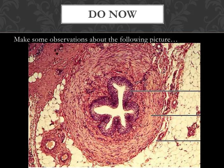
Do Now. Make some observations about the following picture…. Tissues. Chapter 5. objectives. 1. Explain the 4 types of tissues. 2. Identify the different types of epithelial tissue. 3. List the locations of each type of epithelial tissue. . Key terms.

E N D
Do Now • Make some observations about the following picture…
Tissues Chapter 5
objectives • 1. Explain the 4 types of tissues. • 2. Identify the different types of epithelial tissue. • 3. List the locations of each type of epithelial tissue.
Key terms • 1. Histology: The study of tissues • 2. Tissues: Groups of cells which are similar in structure and perform common functions.
Epithelial tissue • Locations: • 1. Covers the body (skin) • 2. Lines cavities, tubes, ducts, blood vessels • 3. Covers organs
Epithelial tissue • Functions: • 1. Protection from physical & chemical injury • 2. Protection against microbial invasion • 3. Contains receptors which respond to stimuli • 4. Filters, secretes & reabsorbsmaterials • 5. Secretes serous fluids to lubricate structures
Epithelium • Facts about Epithelium: • - always have one free surface exposed to the outside or open internal space. • - attached to a basement membrane on the other side. • - tightly packed • - **Avascular- LACK BLOOD VESSELS, rely on diffusion from the underlying connective tissue. • - Divide rapidly!
Classifying epithelium • What characteristics distinguish the following groups of cells?
Classifying epithelium • 1. Layers • a) simple = 1 layer • b) stratified = multiple layers • 2. Shape • a) squamous: thin/flat (think “squished”) • b) columnar: tall slender rectangles (columns) • c) cuboidal: square shaped (like cubes)
Squamous • 1. Simple Squamous • - absorption • Ex: • 2. Stratified Squamous • - physical protection against abrasion • - protects against pathogens/chemicals • Ex:
Squamous • 3. Keratinized Stratified Squamous • - layers of dead squamous cells • - retards water loss • - barrier to organisms
columnar • 1. Simple Columnar • - provides some protection • - absorption/secretion • - usually contain “goblet” cells • Ex: • 2. Stratified Columnar • - *rare • - secretion/absorption • Ex: small portion of male urethra
columnar • 3. Pseudostratified Ciliated Columnar – all cells touch basement membrane Smoking = goodbye cilia
cuboidal • 1. Simple Cuboidal • - secretion/absorption • - secretes sweat • - produces sperm & hormones • - line ducts • Ex: • 2. Stratified Cuboidal • - *rare • - usually only 2-3 layers • - ducts of glands • Ex: Cuboid Cells Duct
Transitional • Stratified • Tolerates repeated stretching • Ex:
Think-pair-share • Identify the types of epithelium on your worksheet by yourself. Go over your answers with the person sitting next to you to see if you got similar answers.
Closing • Take out your cell phones for PollEverywhere!
Do Now • How is epithelial tissue classified? • What is the main function of epithelial tissue? • Which type of epithelium would you find in the intestines that contains goblet cells?
Objectives • Identify slides of tissue using the classification of epitheliail tissue. • Compare and contrast exocrine and endocrine glands. • List and explain the 3 types of exocrine glands.
Identifying Slides • Decide which type of epithelium is being shown on the slide. Try to determine what tissue/organ this is a slide of.
Glandular epithelium • Endocrine: ductless glands (secreted hormones directly into blood stream) • Exocrine: glands contain ducts that connect with surface
Types of glands • Merocrine: release fluid through exocytosis (ex: tears, gastric juices) • Apocrine: release part of cell ( ex: axillary glands, mammary glands) • Holocrine: release and destroy whole cell. (ex: sebaceous glands)
GLands • Types of Glands Animation
Microscope Practice • Please take a slide and a microscope. Go through each of the powers under the microscope, finding the free space that will contain the epithelial tissue. Move the slide accordingly. Call me over to verify that you are looking at the right thing before you move on to the next power.