Cardiac Valve Replacement Surgery
Cardiac Valve Replacement Surgery. Jane Hallam RMH . Anatomy. Indications for Surgery. Aortic Valve

Cardiac Valve Replacement Surgery
E N D
Presentation Transcript
Cardiac Valve Replacement Surgery Jane Hallam RMH
Indications for Surgery • Aortic Valve Stenosis – results from thickening, calcification and/or fusion. Younger pt congenital bicuspid, older pt degenerative changes. Impairment opening, pressure overload, LVH, reduced ventricular compliance. Grading - N 2.5-3.5, mild (AVA >1.5 cm2), moderate (AVA =1.0 – 1.5), severe (AVA <1.0), critical (AVA <0.75) More precise, indexed to pt size, critical AS when AVA Index <0.45cm2
Average survival symptomatic AS 2-3 yrs. Traditional indications presence of angina, CHF, syncope, resuscitation after cardiac arrest. Surgery in asymptomatic controversial. Presence of LV systolic dysfunction, hypotension response exercise, VT or LVH (>15mm), transvalvular peak gradient > 50mmHg are poor prognostic signs, consider early surgery. Pts undergoing CABG’s with AVA <1.1cm2 should have replacement.
Indications for AVR in AS Class I 1 AVR is indicated for symptomatic patients with severe AS.* (Level of Evidence: B) 2 AVR is indicated for patients with severe AS* undergoing coronary artery bypass graft surgery (CABG). (Level of Evidence: C) 3 AVR is indicated for patients with severe AS* undergoing surgery on the aorta or other heart valves. (Level of Evidence: C) 4 AVR is recommended for patients with severe AS* and LV systolic dysfunction (ejection fraction less than 0.50). (Level of Evidence: C) Class IIa AVR is reasonable for patients with moderate AS* undergoing CABG or surgery on the aorta or other heart valves (see Section 3.7 on combined multiple valve disease and Section 10.4 on AVR in patients undergoing CABG). (Level of Evidence: B) Class IIb 1 AVR may be considered for asymptomatic patients with severe AS* and abnormal response to exercise (e.g., development of symptoms or asymptomatic hypotension). (Level of Evidence: C) 2 AVR may be considered for adults with severe asymptomatic AS* if there is a high likelihood of rapid progression (age, calcification, and CAD) or if surgery might be delayed at the time of symptom onset. (Level of Evidence: C) 3 AVR may be considered in patients undergoing CABG who have mild AS* when there is evidence, such as moderate to severe valve calcification, that progression may be rapid. (Level of Evidence: C) 4 AVR may be considered for asymptomatic patients with extremely severe AS (aortic valve area less than 0.6 cm2, mean gradient greater than 60 mm Hg, and jet velocity greater than 5.0 m per second) when the patient’s expected operative mortality is 1.0% or less. (Level of Evidence: C) Class III AVR is not useful for the prevention of sudden death in asymptomatic patients with AS who have none of the findings listed under the class IIa/IIb recommendations. (Level of Evidence: B)
Aortic Valve • Regurgitation Results from abN in AV leaflets such as post inflammatory changes, bicuspid valve, damage from endocarditis, or aortic root dilatation preventing coaptation. Acute AR fr endocarditis or aortic dissection produces LV failure, pul edema as ventricale unable to dilate to handle fluid overload. Chronic AR pressure and volume overload of LV, with progressive dilatation, wall stress, hypertrophy, and symptoms left heart failure. Increased stroke volume will increase pulse pressure, increase SBP and evidence of hyperdynamic circulation.
Class I 1 AVR is indicated for symptomatic patients with severe AR irrespective of LV systolic function. (Level of Evidence: B) 2 AVR is indicated for asymptomatic patients with chronic severe AR and LV systolic dysfunction (ejection fraction 0.50 or less) at rest. (Level of Evidence: B) 3 AVR is indicated for patients with chronic severe AR while undergoing CABG or surgery on the aorta or other heart valves. (Level of Evidence: C) Class IIa AVR is reasonable for asymptomatic patients with severe AR with normal LV systolic function (ejection fraction greater than 0.50) but with severe LV dilatation (end-diastolic dimension greater than 75 mm or end-systolic dimension greater than 55 mm).* (Level of Evidence: B) Class IIb 1 AVR may be considered in patients with moderate AR while undergoing surgery on the ascending aorta. (Level of Evidence: C) 2 AVR may be considered in patients with moderate AR while undergoing CABG. (Level of Evidence: C) 3 AVR may be considered for asymptomatic patients with severe AR and normal LV systolic function at rest (ejection fraction greater than 0.50) when the degree of LV dilatation exceeds an end-diastolic dimension of 70 mm or end-systolic dimension of 50 mm, when there is evidence of progressive LV dilatation, declining exercise tolerance, or abnormal hemodynamic responses to exercise.* (Level of Evidence: C) Class III AVR is not indicated for asymptomatic patients with mild, moderate, or severe AR and normal LV systolic function at rest (ejection fraction greater than 0.50) when degree of dilatation is not moderate or severe (end-diastolic dimension less than 70 mm, end-systolic dimension less than 50 mm).*(Level of Evidence: B)
Mitral Valve • Stenosis MS is an obstruction to LV inflow at the level of the MV as a result of a structural abnormality of the MV apparatus, which prevents proper opening during diastolic filling of the left ventricle. The predominant cause of MS is rheumatic carditis. In patients with MS due to rheumatic fever, the pathological process causes leaflet thickening and calcification, commissural fusion, chordal fusion, or a combination of these processes. Acquired causes of MV obstruction, other than rheumatic heart disease, are rare. These include left atrial myxoma, ball valve thrombus, mucopolysaccharidosis, and severe annular calcification. The normal MV area is 4.0 to 5.0 cm2. Narrowing of the valve area to less than 2.5 cm2 typically occurs before the development of symptoms. MV area greater than 1.5 cm2 usually does not produce symptoms at rest. However, if there is an increase in transmitral flow or a decrease in the diastolic filling period, there will be a rise in left atrial pressure and development of symptoms. The first symptoms of dyspnea in patients with mild MS are usually precipitated by exercise, emotional stress, infection, pregnancy, or atrial fibrillation with a rapid ventricular response. As the obstruction across the MV increases, decreasing effort tolerance occurs.
Although MS is best described as a disease continuum, and there is no single value that defines severity, for these guidelines, MS severity is based on a variety of hemodynamic and natural history data using mean gradient, pulmonary artery systolic pressure, and valve area as follows: Mild (area greater than 1.5 cm2, mean gradient less than 5 mm Hg, or pulmonary artery systolic pressure less than 30 mm Hg), Moderate (area 1.0 to 1.5 cm2, mean gradient 5 to 10 mm Hg, or pulmonary artery systolic pressure 30 to 50 mm Hg), Severe (area less than 1.0 cm2, mean gradient greater than 10 mm Hg, or pulmonary artery systolic pressure greater than 50 mm Hg).
Indications for Surgery for Mitral Stenosis Class I 1 MV surgery (repair if possible) is indicated in patients with symptomatic (NYHA functional class III–IV) moderate or severe MS* when 1) percutaneous mitral balloon valvotomy is unavailable, 2) percutaneous mitral balloon valvotomy is contraindicated because of left atrial thrombus despite anticoagulation or because concomitant moderate to severe MR is present, or 3) the valve morphology is not favorable for percutaneous mitral balloon valvotomy in a patient with acceptable operative risk. (Level of Evidence: B) 2 Symptomatic patients with moderate to severe MS* who also have moderate to severe MR should receive MV replacement, unless valve repair is possible at the time of surgery. (Level of Evidence: C) Class IIa MV replacement is reasonable for patients with severe MS* and severe pulmonary hypertension (pulmonary artery systolic pressure greater than 60) with NYHA functional class I–II symptoms who are not considered candidates for percutaneous mitral balloon valvotomy or surgical MV repair. (Level of Evidence: C) Class IIb MV repair may be considered for asymptomatic patients with moderate or severe MS* who have had recurrent embolic events while receiving adequate anticoagulation and who have valve morphology favorable for repair. (Level of Evidence: C) Class III 1 MV repair for MS is not indicated for patients with mild MS. (Level of Evidence: C)
Mitral Regurge • Common causes of organic MR include rheumatic heart disease, CAD, infective endocarditis, certain drugs, and collagen vascular disease. • MR may also occur secondary to a dilated annulus from dilatation of the left ventricle. • In some cases, such as ruptured chordae tendineae, ruptured papillary muscle, or infective endocarditis, MR may be acute and severe, resulting in cardiogenic shock and acute pul edema. Alternatively, MR may worsen gradually over a prolonged period of time with LV dysfunction, dilatation and ^filling pressures. These 2 ends of the spectrum have quite different clinical presentations. • Ischaemic MR chronic or acute.
Three different MV operations are currently used for correction of MR: 1) MV repair; 2) MV replacement with preservation of part or all of the mitral apparatus; and 3) MV replacement with removal of the mitral apparatus.
Indications for Mitral Valve Operation Class I 1 MV surgery is recommended for the symptomatic patient with acute severe MR.* (Level of Evidence: B) 2 MV surgery is beneficial for patients with chronic severe MR* and NYHA functional class II, III, or IV symptoms in the absence of severe LV dysfunction (severe LV dysfunction is defined as ejection fraction less than 0.30) and/or end-systolic dimension greater than 55 mm. (Level of Evidence: B) 3 MV surgery is beneficial for asymptomatic patients with chronic severe MR* and mild to moderate LV dysfunction, ejection fraction 0.30 to 0.60, and/or end-systolic dimension greater than or equal to 40 mm. (Level of Evidence: B) 4 MV repair is recommended over MV replacement in the majority of patients with severe chronic MR* who require surgery, and patients should be referred to surgical centers experienced in MV repair. (Level of Evidence: C) Class IIa 1 MV repair is reasonable in experienced surgical centers for asymptomatic patients with chronic severe MR* with preserved LV function (ejection fraction greater than 0.60 and end-systolic dimension less than 40 mm) in whom the likelihood of successful repair without residual MR is greater than 90%. (Level of Evidence: B) 2 MV surgery is reasonable for asymptomatic patients with chronic severe MR,* preserved LV function, and new onset of atrial fibrillation. (Level of Evidence: C) 3 MV surgery is reasonable for asymptomatic patients with chronic severe MR,* preserved LV function, and pulmonary hypertension (pulmonary artery systolic pressure greater than 50 mm Hg at rest or greater than 60 mm Hg with exercise). (Level of Evidence: C) 4 MV surgery is reasonable for patients with chronic severe MR* due to a primary abnormality of the mitral apparatus and NYHA functional class III-IV symptoms and severe LV dysfunction (ejection fraction less than 0.30 and/or end-systolic dimension greater than 55 mm) in whom MV repair is highly likely. (Level of Evidence: C) Class IIb MV repair may be considered for patients with chronic severe secondary MR* due to severe LV dysfunction (ejection fraction less than 0.30) who have persistent NYHA functional class III-IV symptoms despite optimal therapy for heart failure, including biventricular pacing. (Level of Evidence: C) Class III 1 MV surgery is not indicated for asymptomatic patients with MR and preserved LV function (ejection fraction greater than 0.60 and end-systolic dimension less than 40 mm) in whom significant doubt about the feasibility of repair exists. (Level of Evidence: C) 2 Isolated MV surgery is not indicated for patients with mild or moderate MR. (Level of Evidence: C)
Tricuspid valve disease • Tricuspid valve dysfunction can occur with normal or abnormal valves. When normal tricuspid valves develop dysfunction, the resulting hemodynamic abnormality is almost always pure regurgitation. This occurs with elevation of RV systolic and/or diastolic pressure, RV cavity enlargement, and tricuspid annular dilatation; RV systolic hypertension occurs in MS, pulmonic valve stenosis, and the various causes of pulmonary hypertension. RV diastolic hypertension occurs in dilated cardiomyopathy, RV infarction, and RV failure of any cause. Pacemaker-induced severe TR is rare but may require intervention. • Abnormalities of the tricuspid valve leading to TR can occur with rheumatic valvulitis, infective endocarditis, carcinoid, rheumatoid arthritis, radiation therapy, trauma (such as repeated endomyocardial biopsies), Marfan syndrome, tricuspid valve prolapse, tricuspid annular dilatation, or congenital disorders such as Ebstein’s anomaly or a cleft tricuspid valve as part of atrioventricular canal malformations. Anorectic drugs may also cause TR. • Tricuspid stenosis is most commonly rheumatic in origin. On very rare occasions, infective endocarditis (with large bulky vegetations), congenital abnormalities, carcinoid, Fabry’s disease, Whipple’s disease, or previous methysergide therapy may be implicated. Right atrial mass lesions represent a nonvalvular cause of obstruction to the tricuspid orifice and may also over time destroy the leaflets and cause regurgitation. Rheumatic tricuspid involvement usually results in both stenosis and regurgitation.
clinical features of tricuspid stenosis include a giant a wave and diminished rate of y descent in the jugular venous pulse, a tricuspid opening snap, and a murmur that is presystolic as well as middiastolic and that increases on inspiration • clinical features of TR include abnormal systolic c and v waves in the jugular venous pulse, a lower left parasternal systolic murmur (holosystolic or less than holosystolic, depending on the severity of hemodynamic derangement) that may increase on inspiration (Carvallo’s sign), a middiastolic murmur in severe regurgitation, and systolic hepatic pulsation. In rare instances, severe TR may produce systolic propulsion of the eyeballs, pulsatile varicose veins, or a venous systolic thrill and murmur in the neck. Other associated clinical features are related to the cause of TR. Moderate or severe TR may be present without the classic clinical features.
Management Class I Tricuspid valve repair is beneficial for severe TR in patients with MV disease requiring MV surgery. (Level of Evidence: B) Class IIa 1 Tricuspid valve replacement or annuloplasty is reasonable for severe primary TR when symptomatic. (Level of Evidence: C) 2 Tricuspid valve replacement is reasonable for severe TR secondary to diseased/abnormal tricuspid valve leaflets not amenable to annuloplasty or repair. (Level of Evidence: C) Class IIb Tricuspid annuloplasty may be considered for less than severe TR in patients undergoing MV surgery when there is pulmonary hypertension or tricuspid annular dilatation. (Level of Evidence: C) Class III 1 Tricuspid valve replacement or annuloplasty is not indicated in asymptomatic patients with TR whose pulmonary artery systolic pressure is less than 60 mm Hg in the presence of a normal MV. (Level of Evidence: C) 2 Tricuspid valve replacement or annuloplasty is not indicated in patients with mild primary TR. (Level of Evidence: C)
Pulmonary Valve • Stenosis Pulmonary valve is the least likely valve to be affected by acquired heart disease, virtually all cases of pulmonary valve stenosis are congenital in origin. Symptoms are unusual in children or adolescents with pulmonary valve stenosis even when severe. Adults with long-standing severe obstruction may have dyspnea and fatigue secondary to an inability to increase cardiac output adequately with exercise. Exertional syncope or light-headedness may occur in the presence of severe pulmonic stenosis with systemic or suprasystemic RV pressures, with decreased preload or dehydration, or with a low systemic vascular resistance state (such as pregnancy). However, sudden death is very unusual. Eventually, with long-standing untreated severe obstruction, TR and RV failure may occur. it appears that congenital mild pulmonary stenosis is a benign disease that rarely progresses, that moderate or severe pulmonary stenosis can be improved with either surgery or balloon valvotomy at very low risk, and that patients who undergo surgery or balloon valvotomy have an excellent prognosis and a low rate of recurrence. Thus, the goal of the clinician is to ascertain the severity of the disease, treat those in whom it is moderate or severe, and infrequently follow up on those with mild disease
Pulmonary Regurgitation • Pulmonary valve regurgitation is an uncommon congenital lesion seen occasionally with what has been described as idiopathic dilation of the pulmonary artery or with connective tissue disorders. In this condition, the annulus of the pulmonary valve dilates, which causes failure of the leaflets to coapt during diastole. • Pulmonary regurgitation also commonly occurs after successful repair of tetralogy of Fallot. • Most physicians would perform pulmonary valve replacement in patients with NYHA class II or III symptoms and severe pulmonary regurgitation, but not for asymptomatic patients.
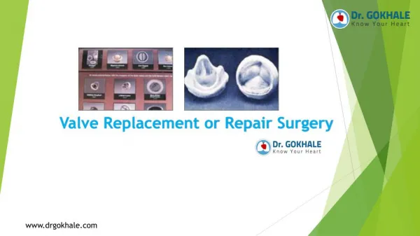
Bioprosthetic/Tissue No lifetime warfarin Less durability Mechanical valve Need for warfarin Better durability Valve types
Need for anticoagulation • Systemic embolization (predominantly cerebrovascular events) occurs at a frequency of approximately 0.7 to 1.0 percent per patient per year in patients with mechanical valves who are treated with warfarin. • In comparison, the risk is 2.2 percent per patient per year with aspirin and 4.0 percent with no anticoagulation. • Patients with mitral valve prostheses are at approximately TWICE the risk as those with aortic valve prostheses