Download
1 / 8
80 likes | 466 Views
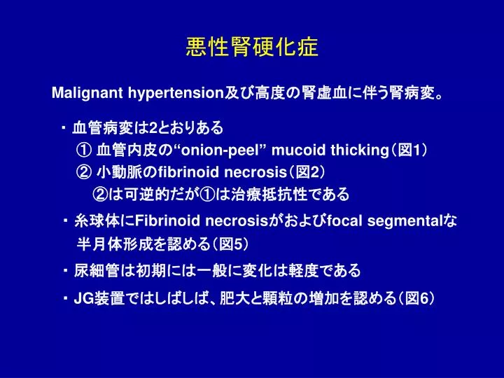
悪性腎硬化症. Malignant hypertension 及び高度の腎虚血に伴う腎病変。 ・ 血管病変は 2 とおりある ① 血管内皮の “onion-peel” mucoid thicking (図 1 ) ② 小動脈の fibrinoid necrosis (図 2 ) ②は可逆的だが①は治療抵抗性である ・ 糸球体に Fibrinoid necrosis がおよび focal segmental な 半月体形成を認める(図 5 ) ・ 尿細管は初期には一般に変化は軽度である

E N D
悪性腎硬化症 Malignant hypertension及び高度の腎虚血に伴う腎病変。 ・ 血管病変は2とおりある ① 血管内皮の“onion-peel” mucoid thicking(図1) ② 小動脈のfibrinoid necrosis(図2) ②は可逆的だが①は治療抵抗性である ・ 糸球体にFibrinoid necrosisがおよびfocal segmentalな 半月体形成を認める(図5) ・ 尿細管は初期には一般に変化は軽度である ・ JG装置ではしばしば、肥大と顆粒の増加を認める(図6)
図1 血管内皮の“onion-peel” mucoid thicking(PAS)
図2 小動脈のFibrinoid necrosis(HE) 血管極 小動脈の フィブリノイド 壊死
図3 小動脈のFibrinoid necrosis(HE) 内皮細胞の 腫大と内皮下 腔の開大に より係蹄腔が よく見えない 血管極小動脈のフィブリノイド壊死(この病変は輸出より輸入動脈でよく認められる)
図4 内皮下腔の拡大(EM) 内皮下腔の 開大を認める 黒矢印は新成 BMを示す
図5 初期の半月体形成 定型的で ない初期の 半月体 係蹄壁のwrinklingがあり初期の虚脱所見
図6 JG装置(PAM) 係蹄壁の Fibirinoid necrosis JG装置の細胞の過形成
解説 • 良性腎硬化症は、人口の高齢化に伴い増加していると思われる。これに対して降圧剤の進歩により現在典型的な悪性高血圧をみることは稀である。 • 悪性腎硬化症で認められる所見は、HUS,TTP,強皮症腎クリーゼ,APS,acute postpartum renal failureでも認められるのでその鑑別のためには臨床情報が重要となる。 • 蛍光抗体法では、良性腎硬化症同様IgMやC3が小動脈壁に陽性を示す。電顕では内皮下にelectron-lucent expansionを認めるのが特徴で時にmesangiolysisを認める。