CSF
Table 1. Subject Information. Introduction

CSF
E N D
Presentation Transcript
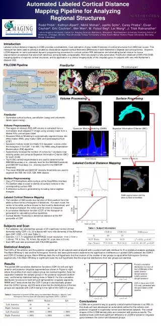
Table 1. Subject Information Introduction Labeled cortical distance mapping (LCDM) provides a probabilistic, local estimation of gray matter thickness of cortical structures from MRI brain scans. This measure has been used in previous studies to characterize regional cortical thickness differences in both Alzheimer’s Disease and schizophrenia. However, LCDM depends on semi-automated dynamic programming procedure for cortical surface ROI delineation and alternating kernel mixture for tissue segmentation procedures that were both laborious and potentially unrepeatable. Here we present a fully-automated, FreeSurfer-initialized LCDM (FSLCDM) mapping pipeline of regional cortical structures, and its application in a clinical imaging study of the cingulate gyrus in subjects with very mild Alzheimer’s disease (AD). FSLCDM Pipeline FreeSurfer FS cortical aparc FS cortical aseg GM Volume Processing Surface Processing WM CSF • FreeSurfer • Generates cortical surface, parcellation (aseg) and volumetric labels (aparc+aseg) • Volume Preprocessing • The region-of-interest (ROI) MR volume is extracted from a normalized, skull-stripped T1 image using a binary mask from a dilated FS's cortical aparc label • Gaussian Mixture Modeling to automatically segment tissue into white matter (WM), gray matter (GM) and cerebralspinal fluid (CSF) • Gaussian mixture model to initially fit 3 Gaussian curves under the histogram (1 for CSF, 1 for GM, 1 for WM) using Expectation-Maximization (EM) algorithm • Systemically increase the number of curves by 1 at places near existing peaks and compute Bayesian Information Criterion (BIC) for each model • Top10 BIC-ranked segmentations are used to determine the WM/GM boundary (i.e., intensity level for the WM/GM threshold), and GM/CSF boundary (i.e., intensity level for the GM/CSF threshold) • The mean WM/GM and GM/CSF intensity thresholds are used to segment the ROI into CSF, GM, WM classes • Surface Preprocessing • Use a FS hemisphere white surface at the Grey/White interface • FS Desikan atlas is used to identify all surface vertices in the corresponding surface ROI • A reference surface is generated by including extra neighbor triangles • Labeled Cortical Distance Mapping • The variation of GM voxels as a function of their position from the white surface forms a histogram of distances: For each voxel, the vertex in the white surface closest to that voxel is determined, and the distance between the vertex and the voxel is measured • A cumulative distribution function (CDF) of the histogram is generated for calculating surface quantities • Cortical Mantle Thickness is defined as distance at the 95th percentile of CDF Gaussian Mixture Modeling (GMM) Bayesian Information Criterion (BIC) Labeled Cortical Distance Mapping CDM Distribution # of GM voxels Voxel (x1 mm) GMM segmentation with the white surface embedded Cortical depth (mm) BIC (lower is better) CSF GM WM Voxel Intensity Number of Peaks (k) • Subjects and Scan • For validation, we selected two groups of 20 cognitively normal (clinical dementia rating CDR = 0), 20 subjects with very mild dementia of the Alzheimer type (DAT, CDR = 0.5) (see Table 1). • Siemens 1.5 T, T1-weighted 3D MPRAGE (voxel resolution: 1mm x 1mm x 1.25mm, TR: 9.7ms, TE: 4.0ms, flip angle:10o, scan time: 6.5 min) • Each MR scan was processed with FSLCDM pipeline. Statistical Analysis The CDFs of the anterior and the posterior cingulate gyri for all subjects were analyzed with a custom-built web interface for R (a statistical analysis package). The Mann Whitney U test and the Kolmogorov Smirnov test were performed to determine the significance of difference in pooled CDFs between the CDR 0 and CDR 0.5 subject groups. Mann-Whitney tests the null hypothesis that the location of the median of two groups is equal while Kolmogorov Smirnov (applied only if the Mann-Whitney is significant) tests the null hypothesis that the empirical distributions from two groups are identical. a. c. CDF Results The pooled GM cumulative distribution functions (CDF) for left and right anterior and posterior cingulate segmented are shown in Figure to right, where the profiles from each subject group are overlaid together. Note the separation between the subject groups in the posterior segments (bottom row), confirmed by statistical testing show in Table 2. For the cumulative distribution functions for both left and right posterior cingulate segments, MWW and T tests show that the CDR 0 group is stochastically greater than the CDR0.5 group, and KS tests show that the distributions of the two groups are separate with CDR 0 being to the right of the CDR 0.5 group. • Pooled cumulative distribution function of the LCDM data of the • left anterior • right anterior • left posterior • right posterior • CDR 0 (blue) and CDR 0.5 (red), generated with the automated LCDM pipeline. Voxel (x1 mm) Voxel (x1 mm) b. d. CDF Table 2: Comparison of pooled LCDMs using one-sided Mann-Whitney U, Welch's t, and Kolmogorov-Smirnov tests with alternative hypothesis CDR0 > CDR0.5. Voxel (x1 mm) Voxel (x1 mm) Conclusions LCDMs are a powerful way to quantify cortical mantle thickness in an ROI. In this study we have developed an automated method for the anterior and posterior cingulate in cognitively normal and demented subjects. The overall shapes of the LCDM density plots are consistent with previous results. The statistical tests confirmed significant difference in LCDM of posterior cingulate gyrus between the control and diseased groups