Reciprocal Lattice
170 likes | 270 Views
Learn about the reciprocal lattice, Brillouin zones, and diffraction theory essential for crystal structure determination in X-ray and neutron scattering. Explore the connection between real and reciprocal space, structure factors, and atomic scattering factors.

Reciprocal Lattice
E N D
Presentation Transcript
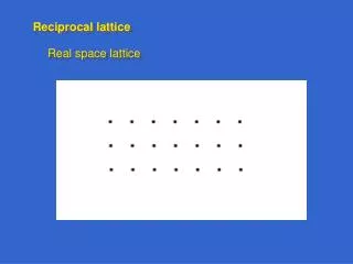
the Reciprocal Lattice the
Ghkl kZB ky kx Brillouin zone The Brillouin zone is the unit cell in reciprocal space (= k-space = momentum space). It is constructed by the Wigner-Seitz method, where k=(000) is the zone center, and the zone boundaries are half way to the nearest reciprocal lattice points: kZB= ½ Ghkl
Brillouin zones for common lattices fcc bcc The reciprocal lattice of fcc is bcc and vice versa. hcp
(-111) (111) (-1-1 1) (020) (220) (000) (200) (11-1) (-1-1-1) (1-1-1) (22-2) (00-2) (20-2) Outer Brillouin zones Reciprocal space can be completely filled with Brillouin zones that are shifted from the central Brillouin zone by reciprocal lattice vectors Ghkl . The reciprocal fcc lattice isbcc. It consists of Ghklwithhkleitherall evenorall odd (units of 2/a). These belong to the two cubic sub-lattices which form the bcc reciprocal lattice (center points and corner points). The edges of the cubes in k-space are 2·2/along, because the spacing of the fcc planes in real space is ½·a(|G|= 2/dplane).
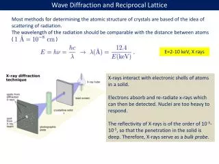
measured intensity k0 k (r) I(k) Real space Reciprocal space General theory of diffraction X-rays scatter off the charge density (r), while neutrons probe the spin density. Diffraction of a coherent plane wave creates aFourier transform of (r) from real to reciprocal space: Ã(k) = (r) eiqr d3rÃ=|Ã|eiI=|Ã|2 = q=kk0 = k-transfer Fourier transform from r to k:Ã(k) = A(r) eikr d3r Inverse transform from k to r:A(k) = (2)3Ã(k) e+ikrd3k
Structure determination for periodic solids • The diffraction pattern is determined by three factors: • The Bragg condition (= energy and momentum conservation) determines the position of the diffraction spots in k-space. It represents the crystal lattice. • The structure factor describes the intensity modulation of the diffraction spots by the atoms inside the unit cell (the basis). This is the quantity measured for protein crystallography. • The atomic scattering factor describes diffraction by the charge distribution inside an individual atom. It is a known quantity. • Large objects in real space correspond to small objects in k-space: • The largest object in real space (the infinite lattice) becomes the smallest object in k-space (a lattice point = -function). • The unit cell represents a medium-sized object in real space. • The smallest object in real space (an atom) modulates the intensity everywhere in k-space by the atomic scattering factor.
Structure factor The structure factor Shkl is given by: Shkl = f exp[-iGhklr] = f exp[-i2 (h·u+k·v+ l·w)] where ris the position of atom inside the unitcell and fits atomic scattering factor. rcan be expressed by integer multiples u,v,w of the real space lattice vectors, just like Gis expressedbytheMillerindices h,k,l in k-space. Ifonechooses a unit cell larger thanthe primitive(= smallest) cell, the structure factor leads to the extinction of certain Bragg spots, because of destructive interference between equivalent atoms in the unit cell. For example, the (100) spot vanishes for the fcc lattice due to the extra face-centered atom at (u,v,w)= (½,0,½).For the diamond lattice in Si both the (100)and (200) spots vanish due to the extra atom at (¼,¼,¼).
Atomic scattering factor The atomic scattering factor fof X-rays is given by: f = (r) · exp[-iGhklr] d3r where is the charge density of a single atom inside the unit cell. The integral over the charge density of an atom is proportional to the number of electrons,i.e. to the atomic number Z. The square ofthe structure factor determines the diffraction intensity. As a result, the diffraction intensity of X-rays increases strongly for heavy atoms (high Z). Light atoms (H,C,…) are hard to detect in the presence of heavy metal atoms. Neutronsscatter very efficiently from light atoms in soft matter,sincethemomentumtransferislargestfor equal masses, such as a H atom and a neutron.
I q½ 1/Rg q Neutron diffraction: Small Angle Neutron Scattering (SANS) Works for light elements (hydrogen, deuterium, soft matter) and for magnetic materials (magnetic moment of the neutron). a Rg Model of a polymer: Rg= Radius of gyration (overall size) aPersistence length (straight sections) Diffracted neutron intensity I plotted versus the k-transfer q
Experimental methods for structure analysis • Energy and momentum conservation imposefour constraintson the diffraction in three dimensions. They cannot all be fulfilled by adjus-ting the three k components of the diffracted wave (with the incident wave fixed). Something else has to give. Either the energy E0 or the wave vector k0of the incident wave needs to be variable. This can be accomplished in several ways: • Use incident x-rays with a continuous energy spectrum (Laue). • Rotate the crystal (popular with protein crystallography). • Use polycrystalline samples (powder diffraction, Debye-Scherrer).
Laue diffraction pattern Laue diffraction pattern of NaCl taken with neutrons. See a projection of k-space.
Powder diffraction pattern Observe rings around the incoming and outgoing beam. (Cylindrical film unfolded.) Extra diffraction rings visible for the ordered Cu3Au alloy. Horizontal scan across the rings for Si powder. The (100), (200) reflections are forbidden in the diamond structure, since their structure factor vanishes.
The phase problem Mathematically, an object in real space can be reconstructed from the ampli-tude of the diffracted wave in k-space by an inverse Fourier transform from k tor. But the amplitude is a complex number of the form A=|A|·ei ,which contains the phase . Only the intensity I=|A|2 is measured, not the phase. Crystallographers have developed tricks to retrieve the phase. In protein crys-tallography, sulfur in selected amino acids can be replaced by selenium. It is chemically similar but heavier. Selenium diffracts X-rays strongly, particularly when the X-ray energy is tuned to an inner shell excitation (anomalous scat-tering). The difference between the diffraction patterns on- and off-resonance provides the phase information. Simple crystal structures can be solved by calculating the diffraction pattern for trial structures containing adjustable parameters. Those are obtained by a least square fitto the diffraction intensities.
Reconstruction of a single nano-object (ptychography) With the advent of laser-like X-ray sources, there has been great interest in Fourier-transforming the diffraction pattern of a single object, for example a virus (next slide). A theorem allows the reconstruction of the phase, as long as the object is located in a known, finite region of space (inside an aperture). The strategy for reconstructing the object uses an iterative method: 1) Start with arbitrary phase in k-space and perform an inverse Fourier trans-form from kto r. The phase error will produce a finite amplitude outside the aperture, where it should be zero. 2) Set the amplitude outside the aperture to zero, but keep the phase inside. 3) Perform a Fourier transform from rto k. The phase error will again give the wrong amplitude, this time in k-space. 4) Reset the amplitude in k-space to the observed diffraction amplitude,but keep the phase. Go back to 1). This loop needs to be iterated many times, but it converges eventually to the correct amplitude and phase in both r and k. Such a method allows lens-less imaging with atomic resolution, limited only by the wavelength of the X-rays.
Diffraction from a single object X-ray diffraction pattern of a single Mimivirus particle imaged at the LCLS at Stanford, which produces laser-like X-rays. The X-ray pulse stripped most of the electrons from the atoms, leading to a Coulomb explosion. But it was so short (< 50 femtoseconds) that the atoms did not have time to move until after this image was obtained (“diffract and destroy”). Combining thousands of such images with various orientations of the virus (tomography) provides a three-dimensional image.