Download
1 / 88
910 likes | 1.08k Views
Learn about the normal hip development, etiology, pathoanatomy, diagnosis, and treatment of developmental dysplasia of the hip (DDH). Understand the risk factors, complications, and imaging modalities. Ideal for medical professionals and students.

E N D
Developmental Dysplasia of the Hip Dr. Nasser ALTurki Teaching assistant @sau . Orthopedics resident @RMH.
Overview • Introduction • Normal Development of the Hip • Etiology and Pathoanatomy • Epidemiology and Diagnosis • Treatment • Complications
Introduction • Developmental Dysplasia of the Hip • DDH - preferred term • Teratogenic hips • Subluxation • Dislocation-usually posterosuperior (reducible vs irreducible) • Dysplasia
Summary • Risk Factors • 1/1,000 born with dislocated hip • 10/10,000 born with subluxation or dysplasia • 80% Female • First born children • Family history (6% one affected child, 12% one affected parent, 36% one child + one parent) • Oligohydramnios • Breech (sustained hamstring forces) • Native Americans (swaddling cultures) • Left 60% (left occiput ant), Right 20%, both 20% • Torticollis or LE deformity
Normal Development • Embryonic • 7th week - acetabulum and hip formed from same mesenchymal cells • 11th week - complete separation between the two • Prox fem ossific nucleus - 4-7 months
Normal Hip • Tight fit of head in acetabulum • Transection of capsule • Still difficult to dislocate • Surface tension
Pathoanatomy • Ranges from mild dysplasia --> frank dislocation • Bony changes • Shallow acetabulum • Typically on acetabular side • Femoral anteversion
Pathoanatomy • Soft tissue changes • Usually secondary to prolonged subluxation or dislocation • Intraarticular • Labrum • Inverted + adherent to capsule (closed reduction with inverted labrum assoc with increased Avascular Necrosis) • Ligamentum teres • Hypertrophied + lengthened • Pulvinar • Fibrofatty tissue migrating into acetabulum
Pathoanatomy • Soft Tissue (Intraarticular) • Transverse acetabular ligament • Contracted • Limbus • Fibrous tissue formed from capsular tissue interposed between everted labrum and acetabular rim • Extraarticular • Tight adductors (adductor longus) • Iliopsoas
Tough Reductions… • Obstacles to reduction • Extraarticular • Tight iliopsoas and adductors • Intraarticular • Labrum • Ligamentum teres • Transverse acetabular ligament • Pulvinar • Redundant capsule (hourglass) • +/- limbus
Etiology and Epidemiology • Multifactorial • Genetics and Syndromes • Ehler’s Danlos • Arthrogryposis • Larsen’s syndrome • Intrauterine environmental factors • Teratogens • Positioning (oligohydramnios) • Neurologic Disorders • Spina Bifida
Diagnosis • Newborn screening • Ortolani’s and Barlow’s maneuvers with a thorough history and physical • Warm, quiet environment with removal of diaper • Head to toe exam to detect any associated conditons (Torticollis, Ligamentous Laxity etc.) • Baseline Neuro and Spine Exam
Diagnosis • Key physical findings • Asymmetry • Limb length- Galeazzi • Abduction ROM • Skin folds • Limp • Waddilng gait / hyperlordosis - bilateral involvement
Ortolani’s Maneuver * After 3 months of age tests become negative
Diagnosis • Some cases still missed • At risk groups should be further screened • AAP • Recs further imaging (e.g. US) if exam is “inconclusive” AND • First degree relative + female • Breech • Positive provocative maneuver (Ortolani or Barlow) • Referral to Orthopaedist
Imaging • X-rays • Femoral head ossification center • 4 -7 months • Ultrasound • Operator dependent • CT • MRI • Arthrograms • Open vs closed reduction
Imaging • Radiographs
Imaging • Radiographs
Imaging • Radiographs
Imaging • Radiographs
Imaging • Acetabular Index
Imaging • Acetabular Index
Imaging • Acetabular Index < 30 wnl
Radiographs Summary • Femoral head appears 4 - 7 months • Shenton’s line • Perkin’s and Hilgenreiner’s lines • Inferomedial quadrant • Center Edge Angle (< 20 abnormal) • Acetabular index • Normal < 30 (Weintroub et al) • Tear drop* • Abnormal widening in DDH *may be only sign in mild subluxation
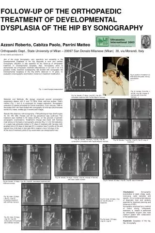
Imaging • Ultrasound • Introduced in 1978 for eval of DDH • Operator dependent • Useful in confirming subluxation, identifying dysplasia of cartilaginous acetabulum, documenting reducibility • Prox Femoral Ossification Center interferes • Requires a window in spica cast (avoid)
Ultrasound Femoral head Abductors Ilium
Ultrasound Femoral head Abductors Ilium
Ultrasound Femoral head Abductors Ilium
Ultrasound Femoral head Abductors Ilium
Ultrasound Graf’s alpha angle
Ultrasound Graf’s alpha angle >60 = normal *line w/ ilium bisects head 50/50
Natural History • Newborn Variable • > 6 months more aggressive tx required due to more extensive pathology and decreased potential for acetabular remodeling • Abnormal Gait, Decreased Abduction and Strength, Increased DJD • Unilateral worse than Bilateral • Subluxation worse than Dysplasia
Treatment Options • Age of patient at presentation • Family factors • Reducibility of hip • Stability after reduction • Amount of acetabular dysplasia
Birth to Six Months • Triple-diaper technique • Prevents hip adduction • “Success” no different in some untreated hips • Pavilk harness (1944) • Experienced staff* • Very successful • Allows free movement within confines of restraints *posterior straps for preventing add. NOT producing abd.
Birth to Six Months • Pavlik harness • Indications • Fully reducible hip* • Child not attempting to stand • Family • Close regular follow-up (every 1-2 weeks) • For imaging and adjustments • Duration • Childs age at hip stability + 3 months
Pavlik Harness • Failures • Poor parent compliance • Improper use by the physician • Inadequate initial reduction • Failure to recognize persistent dislocation • Viere et al 1990 • Bilateral dislocation • Absent Ortolani’s sign • > 7weeks of age
Pavlik Harness • Complications • Avascular necrosis • Forced hip abduction • Safe zone (abd/adduction and flexion/extension) • Femoral nerve palsy • Hyperflexion *Be aware of Pavlik Harness Disease *Follow until skeletal maturity
Birth - Six months • Closed reduction + Spica • Failure after 3 weeks of Pavlik trial
Birth - Six months • Closed reduction • General anesthesia • Arthrogram • Safe zone - avoid AVN • +/- adductor tenotomy • Open reduction if concentric reduction not possible • Usually teratogenic hips in this age group
Open Reduction • Medial approach • Pectineus / adductor longus + brevis • Cannot address simeoultaneous bony work • Antero -lateral • Smith-peterson • Sartorius / Tensor Fascia lata
6 months - 4 years • Present a more difficult problem • Prolonged dislocation • Contracted soft tissues • 6 - 18 months • Closed reduction +/- adductor tenotomy • Spica in human position of 100 degrees of flexion and about 55 degrees abduction (3 months) • Abduction Orthosis 4 wks full time/4 wks nighttime • Open reduction (if closed fails) • Capsulorraphy • CT scan • Spica for 6 wks followed by PT
6 months - 4 years • 18 months - 4 years • Closed reduction • Reducibile - check arthrogram and medial dye pool • Irreducible - Open reduction • Open redcution • Tight - femoral shortening • Stable - +/- pelvic osteotomy