Download
1 / 70
1.03k likes | 2.77k Views
Anatomy of Nose & Paranasal Sinuses. Dr. Vishal Sharma. Development of nose. Face develops from 5 projections: frontonasal process (ectoderm over forebrain) + 2 maxillary processes + 2 mandibular processes (1 st arch)

E N D
Anatomy of Nose & Paranasal Sinuses Dr. Vishal Sharma
Face develops from 5 projections: frontonasal process (ectoderm over forebrain) + 2 maxillary processes + 2 mandibular processes (1st arch) • An ectodermalolfactory placodedividesfronto-nasal process intomedial nasal & lateral nasal process, which give rise to external nasal skeleton • Olfactory placode deepens into olfactory pit & later olfactory sac which forms nasal cavities • Nasal septum develops from mesodermaltecto-septal expansionfrom Rathke’s pouch
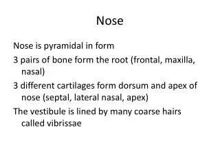
External Framework • Nasal Bone • Frontal Process of Maxilla • Upper Lateral Nasal Cartilage • Lower Lateral Nasal (Alar) Cartilage • Septal Cartilage • Lesser Alar (Sesamoid) Cartilages
Internal Nose • Openings: Anterior nares & posterior nares (Choanae). • Vestibule of nose:lined by skin • Nasal cavity proper: Lateral Wall Medial Wall (Septum) Roof Floor
Nasal Septum 1. Septal cartilage 7. Medial alar crus 2. Ethmoid plate 8. Upper lateral cartilage 3. Vomer 9. Nasal bone crest 4. Palatine crest 10. Frontal spine 5. Maxillary crest 11. Sphenoid rostrum 6. Vomeronasal 12. Membranous septum cartilage
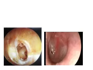
Lateral Nasal Wall 3 - 4 bonyconchaecovered with mucosa to formturbinates.Space below & lateral to turbinate is calledmeatus. Middle meatus contains a round bulla ethmoidalis separated from uncinate process by hiatus semilunaris that leads to a funnel shaped ethmoidal infundibulum.
Meatal Drainage 1. Inferior meatus: Nasolacrimal duct ( guarded at its end by a mucosal valve called HASNER’s valve ) 2. Middle meatus: a. Frontal b. Anterior ethmoid c. Maxillary (natural + accessory ostia) 3. Superior meatus: Posterior ethmoid 4. Spheno-ethmoidal recess: Sphenoid
Ostio-meatal Complex Complex micro-architectural pathway in ethmoid labyrinth that drains anterior group of paranasal sinuses. Consists of: frontal recess + ethmoid infundibulum + hiatus semilunaris + uncinate process + bulla ethmoidalis + middle meatus. O.M.C. pathology leads to infection of all anterior paranasal sinuses (Naumann).
Variants in O.M.C. • Concha bullosa (pneumatized M.T.) • Paradoxically curved middle turbinate • Medially turned uncinate process • Large bulla ethmoidalis • Haller’s cell in orbital floor • Agger nasi cell anterior to M.T. • Onodi Cell with dehiscent Optic Nerve • Mucosal pathology
Blood Supply • Sphenopalatine artery (main artery) • Greater palatine artery • Superior labial artery • Anterior Ethmoidal artery • Posterior Ethmoidal artery 1, 2, 3 & 4 form Kiesselbach’s plexus over Little’s area on anterior septum
Venous drainage Ethmoidal veins:ophthalmic veins cavernous sinus Sphenopalatine vein:pterygoid plexus maxillary vein Woodruff’s venous plexus: present on lateral wall near posterior end of middle turbinate Retro-columellar vein:behind columella
Sensory Nerve Supply • Long & short Nasopalatine nerves • Greater palatine nerve • Infra-orbital nerve branches • Anterior ethmoidal nerve • Olfactory nerve