Download
1 / 1
10 likes | 210 Views
Previous hamstring strain injury reduces knee flexor strength and biceps femoris activation Opar, David 1# ; Dear, Nuala 1 ; Timmins, Ryan 1 ; Williams, Morgan 2; Shield, Anthony 1

E N D
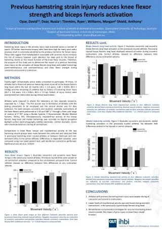
Previous hamstring strain injury reduces knee flexor strength and biceps femoris activation Opar, David1#;Dear, Nuala1; Timmins, Ryan1; Williams, Morgan2; Shield, Anthony1 1 School of Exercise and Nutrition Sciences and Institute of Health &Biomedical Innovation, Queensland University of Technology, Australia 2Division of Sport and Science, University of Glamorgan, Wales #Corresponding author: d.opar@qut.edu.au INTRODUCTION Hamstring strain injury is the primary injury type sustained across a number of sports. Of further importance reinjury rates have been high for many years and a previous insult is commonly identified as the primary risk factor for future injury. These data suggest that maladaptation associated with previous injury increases the risk of reinjury, however scant attention has been paid to the impact of hamstring strains on the neural function of the knee flexor muscles. Therefore, the purpose of this study was to determine the impact of a previous hamstring strain injury on the activation of biceps femoris long head and medial hamstrings (semimembranosus and semitendinosus) and knee flexor strength during concentric and eccentric contraction. METHODS • Twenty-eight recreationally active males consented to participate. Of these, 13 athletes had a history of previous hamstring strain injury all to the biceps femoris long head within the last 18 months (26.2 ± 5.8 years; 1.80 ± 0.04m; 83.0 ± 14.8kg) and the remaining 15 athletes had no history of hamstring strain injury (26.7 ± 5.8 years; 1.8 ± 0.05m; 83.5 ± 7.9 kg). Details of injury history were ascertained from notes taken during clinical examination. • Athletes were required to attend the laboratory on two seperate occasions, seperated by > 7 days. The first session was to familiarise all athletes with the testing procedures to be employed and the second session was for data collection. For both sessions concentric and eccentric isokinetic contractions of the knee flexors were performed at ± 60 and 180°.s-1 through a knee joint range of motion of 5°-90° using a Biodex Systems 3 Dynamometer (Biodex Medical Systems, Shirley, NY). Simulataneously, myoelectrical acitivty of the biceps femoris long head and medial hamstrings was recorded via bipolar pre-gelled Ag/AgCl surface electromyography (sEMG) electrodes (10mm diameter, 25mm inter-electrode distance), during all contractions. • Comparisons in knee flexor torque and myoelectrical activity of the two hamstring muscle groups were made between the uninjured and uninjured limb in previously hamstring strain injured athletes or between dominant and non-dominant limbs in the control athletes. Differences in dependent variables were compared using one tailed paired t-test, with Bonferroni corrections performed. Significance was set at p < 0.0125. RESULTS cont. Biceps femoris long head activity: Figure 2 illustrates concentric and eccentric biceps femoris long head activation in the previously injured athletes. Previously injured biceps femoris long head displayed reduced activation during eccentric contractions only. Control athletes showed no difference between limb differences at any contraction velocity. Figure 2: Biceps femoris long head myoelectrical activity at four different isokinetic velocities derived from previously hamstring strained injured athletes. Negative movement velocities are indicative of eccentric contractions and positive velocities indicate concentric contractions. Error bars display 95% confidence intervals. * p < 0.0125 injured vs uninjured limb. Medial hamstring activity: Figure 3 illustrates concentric and eccentric medial hamstring activation in the previously injured athletes. No between limb differences existed in the injured or control subjects. RESULTS Knee flexor torque: Figure 1 illustrates concentric and eccentric knee flexor torque in the previously injured athletes. Previously injured limbs were weaker at all contraction velocities compared to the contralateral uninjured limb. Control athletes showed no difference between limb differences at any contraction velocity. Figure 3: Medial hamstring myoelectrical activity at four different isokinetic velocities derived from previously hamstring strained injured athletes. Negative movement velocities are indicative of eccentric contractions and positive velocities indicate concentric contractions. Error bars display 95% confidence intervals. CONCLUSIONS Athletes with previous hamstring strain injury were weaker during all concentric and eccentric contractions. Lower levels of myoelectrical activity was only found during eccentric contractions in the previously injured biceps femoris long head. Rehabilitation and prevention strategies for recurrent hamstring strains should consider the impact of prior injury on knee flexor neural function. Figure 1: Knee flexor peak torque at four different isokinetic velocities derived from previously hamstring strained injured athletes. Negative movement velocities are indicative of eccentric contractions and positive velocities indicate concentric contractions. Error bars display 95% confidence intervals. * p < 0.0125 injured vs uninjured limb.