Download
1 / 24
250 likes | 488 Views
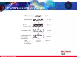
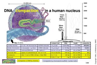
Compaction by chromosome scaffold / nuclear matrix. 10,000 nm. DNA compaction in a human nucleus. 11 nm. 30nm. 1bp (0.3nm). Compaction of DNA by histones. H1. HISTONES are highly conserved, small, basic proteins. Linker histone. H2A. H2B. helix. Histone acetylation

E N D
Compaction by chromosome scaffold / nuclear matrix 10,000 nm DNA compactioninahuman nucleus 11 nm 30nm 1bp (0.3nm) Compaction of DNA by histones JHW
H1 HISTONESare highly conserved,small, basic proteins Linker histone H2A H2B helix • Histone acetylation • is a reversible modification • of lysines in the N-termini • of the core histones. • Result: • reduced binding to DNA • destabilization of chromatin Core histones variable H3 H4 conserved N JHW
Core Histones The histone-fold • The basic structure of ALL • core histones is the same: • 1 long hydrophobic alpha-helix, • bordered by • 2 short hydrophobic alpha helices • that form pairs • H2A - H2B and H3 - H4 • which interact. References:Moudrianakis et al. PNAS 88, 10138 (1991); PNAS 90, 10489 (1993); PNAS 92, 11170 (1995) JHW
Histone octamer assembly Histone octamer H3-H4 tetramer H2A-H2B dimer JHW
H4 H3 H2B H2A Nucleosome features • 146-149 bp DNA in a 1.65 turns of a flat, left-handed superhelix • one pseudo twofold axis centered at the “dyad” (reference: 0 helical turns) • one base-pair precisely at the dyad • sharp bends at + 1.5 and + 4-5 turns • Histone-fold domains organize 121 bp of DNA. The DNA is bound at 10 bp intervals through many contacts, including penetration of arginines at all 14 minor grooves facing the protein core • The grooves from neighboring DNA turns line up; forming channels • H3 and H2B N-termini exit one of these channels every 20bp. • The H4 tail establishes contacts with the next core particle. JHW
1 mM 5 mM Zhou, Gerchman, Ramakrishnan, Travers, Muyldermans Nature 395, 402 (1998) An, Leuba, van Holde, Zlatanova PNAS 95, 3396 (1998) Chromatosome C Core histone octamer + 1 Linker Histone + 2 full turns of DNA (168 bp) N H1 H3 H3 Linker Histone and histone termini control linker DNA entry/exit of chromatosome in chromatin fiber. JHW
Chromatinfibers 11 nm (beads) 30 nm chromatin fiber highly acetylated core histones (especially H3 and H4) + charged N termini (bind DNA on neigboring nucleosomes) • HIGH level of histone H1 • Reduced level of histone H1 • Gene transcription possible • NO gene transcription JHW
Alternative chromatin fiber models Zigzag fiber
Histone modifications 27 Cell (2002) 111, 285-91
5 16 20 8 12 Ac or Me Ac Ac Ac Ac Ac-S-G-R-G-K-G-G-K-G-L-G-K-G-G-A-K-R-H-R-K-V-L-R-D- + + + + + + + + + + 27 4 18 9 23 14 Ac or Me Me Ac Ac Ac Ac A-R-T-K-Q-T-A-R-K-S-T-G-G-K-A-P-R-K-Q-L-A-T-K-A-A-R-K-S-A-P- + + + + + + + + + - N - O O - - C C C g e - N+ P P - a d b C C C - O O O - - - - - - - N P - - C C N C C e - - - C C C C - O - - O - Acetylation of conserved lysines The N-termini of histones H4 and H3, and their acetylation patterns, are absolutely conserved. H4 N-terminus H3 N-terminus DNA backbone binding Lysine Acetyl-CoA HAT (Histone Acetyl-Transferase) Histone Deacetylase reversible reactions CoA e-N-Acetyl-Lysine no DNA binding JHW
(Ac-Lys) antibody nucleosome ppt DNA loop domain H A = -globin genes: domain boundary domain boundary 0 10 30 kb 20 Control: inactive gene DNase I hyper-sensitive site chicken ovalbumin General DNase I sensitivity DNA remaining 0 1 2 DNase I DNase I (U/ml) Acetylation of Chromatin Domains: analysis by the technique of Chromatin Immunoprecipitation (ChIP) Example: the chicken -globin gene domain • High levels of chromatin acetylation, across complete chromatin domains (DNA loops), induces chromatin changes detected as “general DNase I sensitivity” • Within these chromatin domains, at functional genes or transcription factors, • the chromatin structure is interrupted by small “DNase I hypersensitive sites” Hebbes, Clayton, Thorne, Crane-RobinsonEMBO J. 13, 1823 (1994) JHW
TH TH ACGGTC TACCCG ADA2 ADA3 p300 CPB HAT GCN5 HAT HAT P/CAF pol.II TATA Acetylation at Promoters leads to transcriptional activation Thyroid Hormone Receptor example of DNA-binding Transcription Factor + Transcriptional ACTIVATION hormone co-activator Histone Acetyl Transferases HAT TAF 250 II Hyper-acetylated decondensed Chromatin fiber TBP JHW
TACCCG N-CoR Sin3 RPD3 TATA Deacetylation at Promoters leads to Transcriptional repression Transcriptional REPRESSION Without Thyroid Hormone co-repressor Histone Deacetylase HAT X TAF 250 II Hypo-acetylated condensed Chromatin fiber TBP JHW
Acétylation vs déacetylation des histones Chromatine décondensée: Etat transcriptionnel actif Acétylation des histones Chromatine condensée: Etat transcriptionnel réprimé Déacetylation des histones
Histone Methylation C C C C C C Histones can be methylated at lysines or arginines. Example: H3 K4 methylation N Lysine C C C g e N+ a d b C C C O S-adenosylmethyionine HMT (Histone Methyl-Transferase) Histone demethylase DNA backbone binding may not be strongly affected, but specific proteins may recognize these modifications N e-N-monomethyl-Lysine N+ C C C e C C C O S-adenosylmethyionine HMT (Histone Methyl-Transferase) Histone demethylase N Histone demethylases found for K4, K9, K27 and K36 e-N-dimethyl-Lysine N+ C C C e e S-adenosylmethyionine C C C O HMT (Histone Methyl-Transferase) Histone demethylase N N+ C C C e-N-trimethyl-Lysine e e C C C O
Histone methylation : Histone methyltransferases Euchromatin methylation, PcG-dependent gene silencing Enhancer of Zeste Enhancer of Zeste H3 K27 Euchromatin methylation, gene activation trxG-dependent gene silencing Dm Trx, Ash1 Mammalian MLL1-3
Histone demethylases 2 classes: LSD1 JmjC domain proteins
Histone demethylases LSD1 demethylases target mono or dimethyl K4 and K9 of histone H3, depending on their interacting cofactor
Histone demethylases JmjC domain demethylases target many different lysines and are conserved
Histone methylation A reversible phenomenon with many dedicated components Utx JMJD3