Download
1 / 22
230 likes | 378 Views
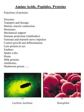
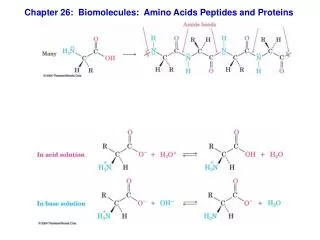
Biomolecules Survey Part 3: Amino Acids, Peptides, and Proteins Lecture Supplement page 238. Repeating unit. Why Bother With Protein Structure?. Molecular structure controls function. Enzyme selectivity Drug design Many others Fundamental protein structure = amide polymer.

E N D
Biomolecules Survey Part 3:Amino Acids, Peptides, and ProteinsLecture Supplement page 238
Repeating unit Why Bother With Protein Structure? Molecular structure controls function • Enzyme selectivity • Drug design • Many others • Fundamental protein structure = amide polymer
Amino AcidsThe Fundamental Building Block of Peptides and Proteins • All amino acids have amine and carboxylic acid groups • All are primary amines (R-NH2) except proline a-carbon • Side chains (R) vary • 18 are S, 1 is R, 1 is achiral Amine (base) + carboxylic acid = proton transfer possible: Keq > 1 at physiological pH Neutral (unionized) form Zwitterionic (ionized) form
Amino Acids • The 20 standard amino acids categorized by side chain properties: • Hydrophilic versus hydrophobic • Hydrophobic nonacidic side chains • Acidic versus basic versus neither (nonacidic)
Amino Acids Hydrophobic acidic side chainsSide chain more acidic than water Tyrosine (Tyr) Cysteine (Cys) Hydrophilic nonacidic side chains Serine (Ser) Threonine (Thr) Asparagine (Asp) Glutamine (Gln)
Amino Acids Hydrophilic acidic side chains Aspartic acid (Asp) Glutamic acid (Glu) Hydrophilic basic side chainsNitrogen lone pairs to accept a proton Lysine (Lys) Arginine (Arg) Histidine (His) Do I have to memorize amino acid structures?
Ala Ser Amino Acids Form Peptides Amino acids link via peptide bond (an amide); form chains Val -2 H2O Serine side chain configuration? Verify with model of complete tripeptide
Amino Acids Form Peptides Ala Ser Val N-terminus C-terminus • A tripeptide (three amino acids) • Naming: Val-Ser-Ala or Ala-Ser-Val? N-terminus C-terminus • Amino acid sequence = primary structure of peptide or protein • Like amino acids, peptides and proteins also have zwitterionic forms:
is planar How Does Peptide Bond Influence Structure? Trans Amino acid chain opposite sides of C-N bond Cis Amino acid chain same side of C-N bond • Torsional strain: Trans < cis; equilibrium favors trans isomer by ~ 2 kcal mol-1 • Amide is conjugated: Conjugation effects: Barrier to rotation around C-N bond ~16 kcal mol-1
The Protein Conformation Problem 3 staggered trans or cis Consider major conformational isomers of a glycine peptide: 3 staggered • Each glycine has 2 x 3 x 3 = 18 major conformations Verify with models • A small protein consisting of 14 glycine has 1814 = 3.7 x 10¹⁷ major conformations! • Number of conformations significantly if more amino acids, or side chains present • Problem: Protein function requires well-organized and restricted structure • Solutions: • Local conformational restrictions: Cis/trans isomers and planarity • Intramolecular hydrogen bonds • Reduced protein flexibility • Reduced structure randomness Results:
Secondary Structure • Structural randomness reduced by intramolecular hydrogen bonds • Causes three basic motifs: The secondary structures of proteins • a-Helix • Clockwise spiral down • H-bonds parallel to axis • Side chains point out from center • Elastic coil: Thinkbook binding There is an H-bond between C=O and N-H of residue 1 and residue 4 (residue 2 and residue 5) (… etc.)
Secondary Structure b-Strand: A “fully extended” polypeptide chain (as opposed to being in a helix) b-Sheet: Two or more aligned, H-bonded b-strands C-terminus N-terminus C-terminus N-terminus • Parallel(N-termini same end) or antiparallel (N-termini opposite ends) • The illustrated b-sheet is antiparallel • b-Sheet more rigid/less elastic than a-helix • Significant component of keratin (hair, wool) and silk • Make your own silk: Thinkbook Appendix C
Secondary Structure (Random) Coil: Not really random, just hard to describe • Key point: Random coils do not have catalytic activity • Denatured proteins adopt the shape of a random coil
Cys Cys Tertiary Structure Tertiary structure: Three-dimensional atomic positions • Aspects of protein structure determined by side chain composition Response to environment: Side chain orientation depends on environment Disulfide bridges: Form loop within one chain, or bond two separate chains • Found in: • Insulin (3) • Keratin (hair) • Others
Quaternary Structure Quaternary structure: Association of two or more subunits by noncovalent bonds • Subunits = proteins, carbohydrates, coenzymes, etc. • Large surface areas noncovalent forces can be significant magnitude Quaternary structure = four subunits
Four levels of protein structure • Primary structure: amino acid sequence • Secondary structure: alpha helix, beta strand / beta sheets • Tertiary structure: spatial arrangement of amino acid residues and disulfide bonds • Quaternary structure: spatial arrangement of subunits and nature of their interactions
Insulin – Secondary Structure and Tertiary Structure • 3 alpha helices • 1 beta strand To play with an interactive 3D-Model of the insulin monomer: http://www.pdb.org/pdb/101/motm_disscussed_entry.do?id=4ins
Insulin – Quaternary Structure Insulin hexamer (inactive form of insulin; long-term storage in the body)
Helix = fuchsia Sheet = yellow Coil = white Protein Structure Representations • stores O2 in muscle tissue via heme • ~70% a-helix • A globular protein (~spherical shape) Myoglobin Worldwide Protein Data Bank: http://www.wwpdb.org/
Helix = fuchsia Sheet = yellow Coil = white Protein Structure Representations Retinol Binding Protein • Important for vision
Helix = fuchsia Sheet = yellow Coil = white Protein Structure Representations Lactate Dehydrogenase • Quaternary structure = four identical protein subunits • Released in bloodstream by damaged muscles • Indicative of heart damage or failure • Subject of Chem 153L experiments