Download

1 / 21
220 likes | 407 Views
9. DIAGNOSTIC NUCLEAR MEDICINE. 9.1. RADIOPHARMACEUTICALS FOR RADIOISOTOPE IMAGING. Nuclear imaging produces images of the distribution of radionuclide in patients. Method of administration: Injection (appropriate for organ) How to determine distribution Blood volume/flow/organ uptake

E N D
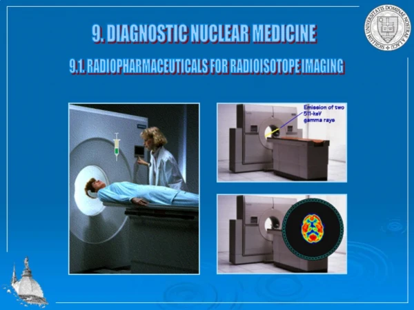
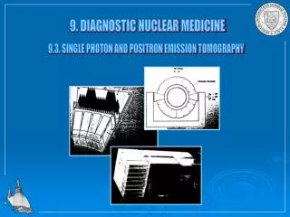
9. DIAGNOSTIC NUCLEAR MEDICINE 9.1. RADIOPHARMACEUTICALS FOR RADIOISOTOPE IMAGING
Nuclear imaging produces images of the distribution of radionuclide in patients. Method of administration: Injection (appropriate for organ) How to determine distribution Blood volume/flow/organ uptake What type of radionuclides? X-rays emitters g-ray emitters (charged particles are absorbed by body tissue) How to detect? Photon detection
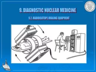
There are several techniques for nuclear imaging: Static planar scintigraphy which provides two-dimensional representations of a three dimensional object by measuring the spatial distribution of the radioisotope in the body, (comparable to a plain X-ray projection). Dynamic planar scintigraphy which measures temporal changes in the spatial distribution of the radioisotopes in the body, by taking multiple images over periods of time which may vary from milliseconds to hours depending on the timescale for the basic function of the organ to be examined. Single photon emission tomography (SPECT) or positron emission tomography (PET) which allows to form three dimensional static or dynamic representations of the organ and organ functions by taking multiple images from different directions.
The great advantage of nuclear medicine is its ability to image qualitatively and quantitatively dynamic physiological processes of different body organs. To highlight a particular organ the radioisotope must be administered in the form of a chemical agent (radiopharmaceutical) which addresses preferably a particular organ (e.g. iodine in thyroid) or the physiological function of a particular organ (e.g. blood flow). Therefore a radiopharmaceutical is typically made of two components, the radionuclide and the chemical compound to which it is bound. Since radiopharmaceuticals are used to study body functions, it is important that they have no pharmacological or toxicological effects which may interfere with the organ function under study. Therefore the pharmaceutical is administered in extremely small amounts (10-9 g).
A number of factors is responsible for the ultimate distribution of the radioisotope: blood flow (percent cardiac input/output of a specific organ) availability of compound to tissue, or the proportion of the tracer that is bound to proteins in the blood basic shape, size, and solubility of molecule which controls its diffusion capabilities through body membranes
The final fate of the radiotracer depends on how the addressed organ deals with the molecule, whether it is absorbed, broken down by intracellular chemical processes or whether it exits from the cells and is removed by kidney or liver processes. These processes determine the biological half-life TBof the radiopharmaceutical (half-life time to reduce material to 50% of its initial amount). To design and administer a radiopharmaceutical with specific localizing properties all these functions as well as the choice of radionuclide has to be taken into account.
The choice of the appropriate radioisotope for nuclear imaging is dictated by the physical characteristics of the radioisotope: a suitable physical half-life; long enough for monitoring the physiological organ functions to be studied, but not too long to avoid long term radiation effects decay via photo emission (X-ray or -ray) to minimize absorptioneffects in body tissue photon must have sufficient energy to penetrate body tissue withminimal attenuation but photon must have sufficiently low energy to be registered efficiently in detector and to allow the efficient use of lead collimator systems (must be absorbed in lead) decay product (daughter) should have minimal short-lived activity
The effective half-life TEof a radiopharmaceutical is a combination of the physical half-life T1/2 and the biological half-life TB : The effective half-life of radiopharmaceuticals containing a long lived radioisotope can be reduced by choosing a chemical component with a short biological half-life. A close matching of the effective half-life with the duration of the study is important for dosimetric considerations. It also is important as far as the availability and expense of the radiopharmaceutical is concerned.
Radioisotopes in common use are: 99Tcm (T1/2=6.02h, E=140 keV) is used in more than 70% of all medical applications 226Ra (T1/2=1600 a, E=186 keV) is an -emitter ('s are absorbed in body tissue), is used for highly localized studies 67Ga (T1/2=78.3h, E =93 keV, 185 keV, 300 keV) is often used as tumor localizing agent (gallium citrate) 123I (T1/2=13h, E=159 keV) bonds good with proteins and molecules that can be iodinated. It has replaced 131I (T1/2=6d, E=364 keV) because of the reduced radiation exposure 81Krm (T1/2=13s, E=190 keV) is a very short-lived gas used to perform lung ventilation studies, (short half-life limits its application)
Important for PET studies are neutron deficient isotopes which decay by positron emission. Positrons annihilate with electrons emitting two E=511 keV photons in opposite direction. Currently used positron emitters are: 68Ga (T1/2=68 m, E= 511 keV) 82Rb (T1/2=1.3 m, E = 511 keV) 18F (T1/2=110 m, E = 511 keV), (used in more than 80% of all PET applications) 13N (T1/2=10 m, E = 511 keV) 11C (T1/2=20.4 m, E = 511 keV) Most of the positron emitters are still being studied in terms of their applicability for diagnostic purposes.
The production of radioisotopes is expensive! It is based on four different methods: nuclear fission (reactor breeding) neutron activation processes charged particle induced reactions radionuclide generator (chemical method) Each method provides useful isotopes with differing characteristics for nuclear imaging.
Nuclear Fission: In nuclear fission, the nuclei of atoms are split, causing energy to be released. • The atomic bomb and nuclear reactors work by fission. The element uranium is the main fuel used to undergo nuclear fission to produce energy since it has many favorable properties. Uranium nuclei can be easily split by shooting neutrons at them. Also, once a uranium nucleus is split, multiple neutrons are released which are used to split other uranium nuclei. This phenomenon is known as a chain reaction.
NUCLEAR FISSIONtakes place in the reactor core and is induced by the slow neutron induced fission (break-up) of 235U into medium mass nuclei. The most common radioisotopes produced by fission (with subsequent isotope separation based on different physical and chemical methods) are 99Mo (which decays to 99Tcm), 131I, and 133Xe!
NEUTRON ACTIVATIONis based on capture reactions of thermal neutrons (produced in the reactor as consequence of the fission process) on stable isotopes which are positioned near the reactor core. Examples for radioisotope production via neutron capture are: • 98Mo + n 99Mo + • 50Cr + n 51Cr + • 31P + n 32P + • 32S + n 32P + p The yield Nrfor the radioisotope production over the time period t depends on the cross section [cm2] of the neutron capture process, the neutron-flux [cm-2s-1], the number of target nuclei nT, and the decay constant = ln2/T1/2
The table shows several radioisotopes produced by neutron absorption. 1 barn = 10-24 cm2 Disadvantage is that the produced radioisotope is typically an isotope of the target element, therefore chemical separation is not possible. This means that the (n,) produced radionuclide are not carrier-free.
CHARGED PARTICLE INDUCED REACTIONSare based on the use of accelerators. Charged particles like protons, deuterons or alphas are accelerated to energies between 1 to 100 MeV and bombard a target material. The most used accelerator type is the cyclotron, where the charged particles are accelerated by oscillating accelerating potentials perpendicular to a deflecting magnetic field.
Examples of typical reactions in the target are listed in the table The advantage of production via charge particle interaction is the large difference in Z between the target material and the radionuclide. That allows good physical and chemical separation procedures.
RADIONUCLIDE GENERATORSallow to separate chemically short-lived radioactive daughter nuclei with good characteristics for medical imaging from long-lived radioactive parent nuclei. Typical techniques used are chromatographic absorption, distillation or phase separation. This method is in particular applied for the separation of the rather short-lived 99Tcm (T1/2=6 h) from the long lived 99Mo (T1/2=2.7 d). Applying the radioactive decay law the growth of activity of the daughter nuclei A2 with respect of the initial activity of the mother nucleus A10can be expressed in terms of their respective decay constants 2 and 2 with 2 >> 1: Milking cow analogy
In a generator such as the Mo-99/Tc-99m radionuclide generator in which the half-life of the mother nuclide is much longer than that of the daughter nuclide, 50% of equilibrium activity is reached within one daughter half-life, 75% within two daughter half-lives. Hence, removing the daughter nuclide from the generator ("milking" the generator) is reasonably done every 6 hours or, at most, twice daily in a Mo-99/Tc-99m generator. Most commercial Mo-99/Tc-99m generators use column chromatography, in which Mo-99 is adsorbed onto alumina. Pulling normal saline through the column of immobilized Mo-99 elutes the soluble Tc-99m, resulting in a saline solution containing the Tc-99m which is then added in an appropriate concentration to the kits to be used. The useful life of a Mo-99/Tc-99m generator is about 3 half lives or approximately one week. Hence, any clinical nuclear medicine units purchase at least one such generator per week or order several in a staggered fashion.