Download
1 / 271
2.71k likes | 2.92k Views
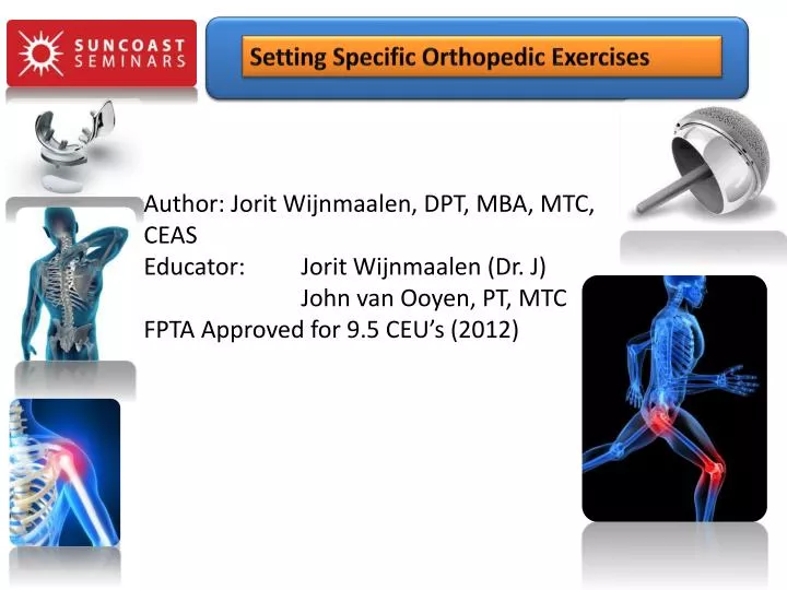
Setting Specific Orthopedic Exercises. Author: Jorit Wijnmaalen , DPT, MBA, MTC, CEAS Educator: Jorit Wijnmaalen (Dr. J) John van Ooyen , PT, MTC FPTA Approved for 9.5 CEU’s (2012). Setting Specific Orthopedic Exercises. About this course:.

E N D
Setting Specific Orthopedic Exercises Author: JoritWijnmaalen, DPT, MBA, MTC, CEASEducator: JoritWijnmaalen (Dr. J) John van Ooyen, PT, MTCFPTA Approved for 9.5 CEU’s (2012)
Setting Specific Orthopedic Exercises About this course: The Setting Specific Orthopedic Exercise Course is the new version of our Anatomy of Exercise course and now includes exercise protocols for most major Orthopedic and Spine Surgeries. We have added more material to the course and it has been submitted for FPTA approval for 9.5 CEU's. This course is required for the CORS certification, but if you have taken The Anatomy of Exercise in 2012 or 2011, you do not need to take this course to become eligible for the CORS certification. This hands-on exercise course will review in depth clinical protocols that are currently in place for the various, common orthopedic procedures including joint replacement, ligamentous and tendon repairs surgeries and many spine surgeries. This clinical review will include protocols as they are applied in the various rehab settings including Inpatient Acute Care, SubacuteRehab and Skilled Nursing settings, Homecare and Outpatient.
SunCoast Seminars Setting Specific Orthopedic Exercises • Why do we need to know and understand the anatomy of muscle? • This will allow the clinician to specifytheir exercise program geared towards the function of the muscle. Different muscles have different functions and these functions are in part defined by the anatomy of the muscle. • There are approximately 639 skeletal muscles in the human body. There are different types of muscles, each with their distinct anatomy. • Understanding the anatomy of the muscle will help the clinician understand how different (intrinsic and extrinsic) factors can impact muscles and exercising. • We are looked upon as the experts when it comes to exercise therapy. Understanding the anatomy of muscles is an important part of being an exercise expert.
Setting Specific Orthopedic Exercises • Program Objectives: • Reviewing muscular anatomy and physiology • This will include a review of tissue healing • Discuss how extrinsic factors such as medication, progression, exercise objectives etc. may affect the exercise therapy program • Discuss how intrinsic factors including disease processes age, vital signs etc. may affect muscles and exercise programs. • Discuss the basics of exercise therapy • Discuss common exercise principles • Open chain vs. closed chain • Eccentricisometricconcentric
SunCoast Seminars Setting Specific Orthopedic Exercises • About the educator: • Background • Education • Work experience • Hobbies
About SunCoast Seminars Setting Specific Orthopedic Exercises • 5 educators • Dr. Brian Healy • Dr. Willem Stegeman • Dr. Jorit Wijnmaalen • John Van Ooyen, PT • Dr. Nathan A. Possert, PT, DPT
About SunCoast Seminars Setting Specific Orthopedic Exercises More courses: • Orthopedic Joint Replacement course: 9.5 CEU • Comprehensive Management of back & neck pain: 9.5 CEU • Clinical Imaging for the Rehab Specialist, 9.5 CEU • Joint Replacement, online: 7 CEU • Thoracic Outlet Syndrome: 6.0 CEU, Online • HIV/Medical Errors/Abuse: 4 CEU • The Anatomy of Excercise: online • An Introduction to Manual Therapy : 9.5 CEU • Setting Specific Orthopedic Exercises : 9.5 CEU’s • CORS: 9.0 CEU’s
SunCoast Seminars Setting Specific Orthopedic Exercises Muscular anatomy and physiology Muscle Types: Smooth muscles Cardiac muscles Skeletal muscles
SunCoast Seminars Setting Specific Orthopedic Exercises • Smooth muscles • These muscles are very important in physiological regulation. • Help to regulate the flow of blood. • Help control BP • They control the movement of food through the digestive system. • Control of the uterus during labor • Contraction of a smooth muscle cell is generated by a sliding mechanism of the myofilaments. • Contraction is involuntary and may be initiated by • Nerve impulse • Hormones (i.e. cardiac function) • Mechanical change to the muscle
SunCoast Seminars Setting Specific Orthopedic Exercises • Smooth Muscles : • Crucial difference with skeletal muscles: nervous control is absolutely required for skeletal muscles, smooth muscles can, to a degree, work without nervous stimulation! • Lastly, these muscles are not striated (the myofilaments are arranged into light and dark bands as in striated muscles). Striations are formed by alternating segments of thick and thin protein filaments, which are anchored by segments called T-lines
SunCoast Seminars Setting Specific Orthopedic Exercises • Cardiac Muscles : • This muscle may look like a skeletal muscles (especially the contraction of it since they are striated as well) but it acts much like smooth muscle (it does not require nervous system input to function) • The attachment site between cells is called an intercalated disc, which is present only in cardiac muscle cells and allows forces to be transmitted from one cell to the next.
SunCoast Seminars Setting Specific Orthopedic Exercises • Skeletal Muscles: • Striated (banded) type. This distinctive banding pattern of striated muscle is an effect that comes from the alignment of sarcomeres in register across the myofibrils • Skeletal muscles are under voluntary control; no skeletal muscle works without “orders” from the nervous system • Skeletal muscles have elongated muscle cells (fibers) with multiple nuclei lying along the periphery of the cell. The sarcoplasmof each cell is contained by a sacrolemma(plasma membrane) and an external lamina. • Each muscle contains many myofibrils and each myofibril contains thin actineand thick myosinmyofilaments. • These muscles normally make up the largest portion of a person's lean body mass
SunCoast Seminars Setting Specific Orthopedic Exercises • Skeletal Muscles • These are the muscles that are responsible for all voluntary movements (movements controlled by the central nervous system and which typically are directed at some sort of interaction with the environment) • These muscles only contract in response to instructions from the central nervous system (with a few exceptions) • In short, skeletal muscles have the following functions: • provide joints with the forces necessary to produce movement • to control movement • to stabilize and protect joints when loads are applied to them. • generating heat, maintaining normal body temperature, because they account for 40% of the body mass.
Setting Specific Orthopedic Exercises • Skeletal Muscles • Skeletal muscles are a striated type of muscle with a rich blood supply, extensive afferent and efferent innervations and an extremely high metabolic capacity. • Skeletal muscles have a tremendous adaptive capacity that allows them to hypertrophy, atrophy, increase in physiological length, decrease in physiological length and change metabolic capacities. • Out of the three muscle types discussed, the skeletal muscle are the muscles that we will be most concerned within this course.
Setting Specific Orthopedic Exercises Muscular anatomy and physiology Let’s review!
Setting Specific Orthopedic Exercises • The Anatomy review of a skeletal muscle • Each muscle cell is surrounded by a basal lamina and connective tissue. • They are bound to each other and to surrounding tissues by connective tissue to form a gross "muscle". Skeletal muscle fibers are NOT joined by cell junctions. • The endomysium consists of the basal lamina and thin connective tissue that surrounds individual muscle cells. • The perimysium consists of sheets of connective tissue which separate the fibers into groups known as fascicles. • The epimysium surrounds the groups of fasicles that comprise the “muscle”.
Setting Specific Orthopedic Exercises • Endomysium – delicate connective tissue sheeth that encloses each muscle fiber • Fasciculus – bundle of muscle fibers covered by perimysium (coarser fibrous membrane) • Epimysium – covers bundle of fasciculi (entire muscle); blends into either: • Tendon – cord of dense, fibrous tissue attaching a muscle to a bone • Aponeurosis – fibrous or membranous sheet connecting a muscle and the part is moves (usually found on torso)
Setting Specific Orthopedic Exercises • The Anatomy review of a skeletal muscle • Connective tissue transmits the mechanical force of muscle. • Tendons connect muscle to bone. The myotendinous junction occurs at the end of the muscle cell where the terminal actin filaments connect to the plasma membrane • Skeletal muscle fibers are multi-nucleatedcells that arise by fusion of mono-nucleatemyoblasts. • The many nuclei are located at the periphery of the cell. • Mono-nucleate satellite cells, associate with the muscle fiber and reside within the muscle basal lamina. They promote limited regeneration of muscle in the adult.
Setting Specific Orthopedic Exercises The Yellow line is corresponding to the tendon. How do we classify this Connective tissue? Dense Regular. The yellow arrows are pointing the nuclei of the fibroblasts making the collagen. The blue line is showing where the Striated Muscle is beginning The muscle-tendon junction
Setting Specific Orthopedic Exercises • Innervation of a Skeletal Muscle • Skeletal muscle is innervated and highly vascularized, due to its high energy requirements. It is penetrated of blood vessels into the epimysium with branches into the peri- and endomysium.
Setting Specific Orthopedic Exercises • Innervation of a Skeletal Muscle • Motor end plates (neuromuscular junctions) are specialized sites at which a nerve contacts a muscle cell. • The terminal branches of motor axons lie in the surface of the muscle cell, where the plasma membrane is highly folded. • Muscle action begins at the motor end plate (or neuromuscular junction), which is analogous to a synapse • Acetylcholine(ACh) binds to receptors localized in the muscle membrane at the motor end plate, resulting in local depolarization at the end plate. • When this depolarization exceeds the threshold, it will result in an action potential
Setting Specific Orthopedic Exercises Neuromuscular Junction or Motor End Plate axon of Motor (Efferent) Neuron White arrow - Skeletal Muscle Fiber
Setting Specific Orthopedic Exercises • Innervation of a Skeletal Muscle • Additional proprioceptor endings (Golgi tendon organs) are located at the point where muscle fibers attach to tendon • These Golgi tendon organs (GTO) respond to tension (force) exerted by the muscle; activity in these axons inhibits muscle contraction (they are for instance stretched when a joint is swollen).
Setting Specific Orthopedic Exercises • Nerve Conduction • Both nerve cells and muscle cells are excitable • Their cell membrane can produce electrochemical impulses and conduct them along the membrane. • In muscle cells, this electric phenomenon is also associated with the contraction of the cell • The origin of the membrane voltage is the same in nerve cells as in muscle cells. In both cell types, the membrane generates an impulse as a consequence of excitation. • The long nerve fiber, the axon, transfers the signal from the cell body to another nerve or to a muscle cell • The axon may be covered with an insulating layer called the myelin sheath, which is formed by Schwann cells
Setting Specific Orthopedic Exercises • Nerve Conduction • This myelin sheath is not continuous but divided into sections, separated at regular intervals by the nodes of Ranvier • The junction between an axon and the next cell with which it communicates is called the synapse. • Information proceeds from the cell body uni-directionally over the synapse, first along the axon and then across the synapse to the next nerve or muscle cell (think about peripheral leasion) • The part of the synapse that is on the side of the axon is called the pre-synaptic terminal • The part on the side of the adjacent cell is called the postsynaptic terminal. Between these terminals, there exists a gap. • A chemical neurotransmitter, released from the pre-synaptic cell, is responsible for the impulse to transfer across the synapse.
Setting Specific Orthopedic Exercises • Nerve Conduction • This transmitter, when released, activates the postsynaptic terminal. The synapse between a motor nerve and the muscle it innervates is called the neuromuscular junction
Setting Specific Orthopedic Exercises • Nerve Conduction • If a nerve cell is stimulated, the trans-membrane voltage necessarily changes. The stimulation may be • excitatory (i.e., depolarizing; characterized by a decrease in the normally negative resting voltage) or • inhibitory (i.e., hyperpolarizing, characterized by an increase in the magnitude of the membrane voltage). • After stimulation the membrane voltage returns to its original resting value • If the excitatory stimulus is strong enough, the trans-membrane potential reaches the threshold, and the membrane produces a characteristic electric impulse, the nerve impulse. • Remember the Na+/K+ pump?
Setting Specific Orthopedic Exercises • Nerve Conduction • Many factors may affect nerve conductivity but discussion of those factors would be outside the scope of this lecture. • Temperature • Properties of the membrane • Sodium levels • Age • Anatomical changes because of disease (ALS)
Setting Specific Orthopedic Exercises • Nerve Conduction • A myelinated axon (surrounded by the myelin sheath) can produce a nerve impulse only at the nodes of Ranvier • In these axons the nerve impulse propagates from one node to another • The myelin sheath increases the conduction velocity • The conduction velocity of the myelinated axon is directly proportional to the diameter of the axon
Setting Specific Orthopedic Exercises Nerve Conduction
Setting Specific Orthopedic Exercises • Types of Skeletal muscles • Not all skeletal muscles are the same. • Some cells are thicker than others • Some shorten faster • Some produce more tension • Some fatigue more rapidly • Looking at these different features, there appear to be three major types of skeletal muscles:
Setting Specific Orthopedic Exercises • Types of Skeletal muscles • Slow Twitch • Fast Fatigue Resistant • Fast Twitch Fatigable
Setting Specific Orthopedic Exercises • Slow Twitch • These muscles produce the least amount of force. They actually produce less than half the force produced by fast twitch fatigue resistant fibers and are most resistant to fatigue. • Slow twitch muscles use oxygen for power and have a predominance of aerobic enzymes. • Slow twitch muscles are red, because they contain lots of blood vessels. • These muscle fibers are "hit", or engorged with nitrogen-rich blood, during higher rep training, specifically in sets of 12 to 20 reps. • Slow twitch muscles are used for holding posture
Setting Specific Orthopedic Exercises • Fast Twitch (Type II) • Fast Twitch fibers use anaerobic metabolism to create fuel and so they are much better at generating short bursts of strength or speed than slow muscles. • These types of muscles are best trained during sets of 2-5 repetitions. • They fatigue more quickly. • Fast twitch fibers generally produce the same amount of force per contraction as slow muscles, but they get their name because they are able to fire more rapidly. • Having more fast twitch fibers can be an asset to a sprinter since she needs to quickly generate a lot of force (genetically determined, 50/50 on average; some research suggests that some fibers might be able to convert).
Setting Specific Orthopedic Exercises • Two Types: • Type IIa Fibers / Fast Fatigue Resistant • These fast twitch muscle fibers are also known as intermediate fast-twitch fibers. • They can use both aerobic and anaerobic metabolism almost equally to create energy. • In this way, they are a combination of Type I and Type II muscle fibers. • Produce forces greater than slow twitch fibers but less than fast twitch fatigable fiber. • These fibers are more resistant to fatigue than fast fatigable but less fatigue resistant than slow twitch fibers.
Setting Specific Orthopedic Exercises • Type IIb Fibers • These fast twitch fibers use anaerobic metabolism to create energy and are the "classic" fast twitch muscle fibers that excel at producing quick, powerful bursts of speed. • This muscle fiber has the highest rate of contraction (rapid firing) of all the muscle fiber types, but it also has a much faster rate of fatigue and can't last as long before it needs rest. • Produce the greatest amount of force • Are least resistant to fatigue • Force produced is typically 2-3 times greater than fast twitch fatigue resistant fibers
Setting Specific Orthopedic Exercises • Low frequency stimulation of motor units of type II fibers transforms these fibers in type I fibers (endurance training, easier to accomplish) • High frequency stimulation of motor units of type I fibers transforms these fibers in type II fibers (strength training, harder to accomplish) This is due to rest periods with low frequent stimulation of type II fibers, only metabolism and muscle fiber diameter stay increased.
Setting Specific Orthopedic Exercises • Conclusion • So the lesson here is quite simple. As we are exercising our patients, we must keep in mind the main objective of our exercise program. • In order to recruit the largest possible number of muscle fibers of both types during the exercise program, we must vary the repetition ranges. • Keeping in mind that on average, there is a 50/50 split of these fibers so… • Any therapist, who puts a patient on an exercise program that doesn't include a variation of repetition ranges might significantly limit the success of the exercise program.
Setting Specific Orthopedic Exercises • Skeletal Muscle Fiber Arrangement • It is important to realize that there are different alignments of muscle fibers in the various skeletal muscles. • These different fiber arrangements will have an effect on the length, mechanical properties and the number of muscle fibers of a muscle. • Muscle fibers can be arranged in parallel or at angles to the tendon. • Parallel fibered muscles are muscle composed of parallel aligned fibers. These muscles have long muscle fibers that can produce a large excursion on the tendon. • Fusiform • Triangular • Spiral • Pinnated fibers muscles are muscles composed of angled fibers • Unipinnate • Bipinnate • Multipinnate
Setting Specific Orthopedic Exercises Structure & Function of a Skeletal muscle
Setting Specific Orthopedic Exercises • The cell comprises a series of striped or striated, thread-like myofibrils. • Within each myofibril there are protein filaments that are anchored by dark Z line. • The fiber is one long continuous thread-like structure. • The smallest cross section of skeletal muscle is called a sarcomere which is the functional unit within the cell. It extends from one Z line to the next attached Z line. The individual sarcomere has alternating thick myosin and thin actin protein filaments. • Myosin forms the center or middle of eache M line. Thinner actin filaments form a zig zag pattern along the anchor points or Z line.
Setting Specific Orthopedic Exercises • Muscle Contraction • Upon stimulation by an action potential, skeletal muscles perform a coordinated contraction by shortening each sarcomere. • The best proposed model for understanding contraction is the sliding filament model of muscle contraction. • Actin and myosinfibers overlap in a contractile motion towards each other. • ATP binds to the cross bridges between myosin heads and actin filaments. The release of energy powers the swiveling of the myosin head • Myosin filaments have club-shaped heads that project toward the actin filaments. • Larger structures along the myosin filament called myosinheads are used to provide attachment points on binding sites for the actin filaments.