Download
1 / 117
1.58k likes | 4.51k Views
Pathology of Cardiovascular System. Dr. S.L. Beh philipbeh@pathology.hku.hk. Overview. Review of basics Ischaemic heart diseases Coronary artery occlusions Myocardial infarction Valvular heart diseases Degenerative valvular diseases Rheumatic heart disease Bacterial endocarditis Shock

E N D
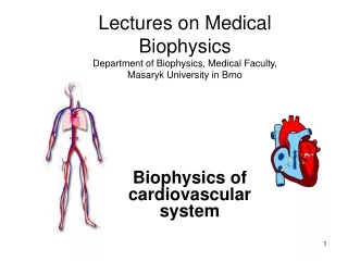
Pathology of Cardiovascular System Dr. S.L. Beh philipbeh@pathology.hku.hk
Overview • Review of basics • Ischaemic heart diseases • Coronary artery occlusions • Myocardial infarction • Valvular heart diseases • Degenerative valvular diseases • Rheumatic heart disease • Bacterial endocarditis • Shock • Hypovoleamic shock • Cardiogenic shock • Septiceamic shock • Anaphylactic shock
Review • Atherosclerosis • Epidemiology of coronary artery disease • Physiology of the cardiac cycle • Anatomy of the myocardium • Vascular supply of the myocardium
Taken from Colour Atlas of Anatomy – Roden, Yokochi and Lutjen-Drecoll
Taken from Colour Atlas of Anatomy – Roden, Yokochi and Lutjen-Drecoll
Taken from Colour Atlas of Anatomy – Roden, Yokochi and Lutjen-Drecoll
Taken from Colour Atlas of Anatomy – Roden, Yokochi and Lutjen-Drecoll
Taken from Colour Atlas of Anatomy – Roden, Yokochi and Lutjen-Drecoll
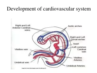
Anatomy of the myocardium • Cardiac muscle cells form a collection of branching and anastamosing striated muscles. They make up 90% of the volume of the myocardium. • Unlike skeletal muscles, they contain ten times more mitochondria per muscle cell. This reflects their extreme dependence on aerobic metabolism. They do not need to rest!!
Vascular supply of the myocardium • Predominant blood supply is from the coronary arteries, which arises from the aorta and runs along an epicardial route before penetrating the myocardium as intramural arteries. Effectively a “one-way street” flow and supply. • Coronary arterial blood flow to the myocardium occurs during ventricular diastole; when the microcirculation in the myocardium is not compressed by cardiac contraction. The “one^way street” only flows within a fixed time span.
Coronary Angiography L = Left main trunk A= Anterior descending C= Circumflex R= Right coronary P=Posterior descending
Areas of supply (perfusion) • The left coronary trunk gives rise to:- • Left Anterior Descending (LAD) and the Left Circumflex (LCX) • Right Coronary Artery (RCA)
Areas of perfusion • Left anterior descending (LAD) – supplies most of the apex of the heart, the anterior wall of the left ventricle and the anterior two-thirds of the ventricular septum. • Left circumflex branch supplies the lateral wall of the left ventricle. • The right coronary artery in 80% of the population supplies the right ventricle, the posterior third of the ventricular septum and the posterior-basal wall of the left ventricle. (Right dominant circulation)
Ischaemic Heart Diseases • This is a generic name for a group of closely related syndromes that result from myocardial ischaemia. • In over 90%, this is due to a reduction in coronary blood flow. (Decrease in supply) • Other conditions arise as a result of increases in demand e.g. hypertrophy, shock, increase heart rate, etc.
Diminished Coronary Perfusion • Fixed coronary obstruction • More than 90% of patients with IHD • One or more lesions that causes at least 75% reduction of the cross-sectional area of at least one of the major epicardial arteries.
Clinical Manifestations • Angina Pectoris • Myocardial Infarction • Chronic ischaemic heart disease • Progressive heart failure consequent to previous myocardial infarction. • Sudden Cardiac Death
Angina Pectoris • This is a symptom complex. Symptoms caused by transient myocardial ischaemia that falls short of inducing the cellular necrosis that defines myocardial infarction. • Three variants:- • Stable angina • Prinzmental angina • Unstable angina
Angina Pectoris • Stable Angina – Most common form. Chronic stenosing coronary atherosclerosis, reaching a critical level, leaving the heart vulnerable to increased demand. • Typically relieved by rest or a vasodilator
Prinzmental Angina • Uncommon pattern • Occurs at rest • Documented to be due to arterial spasm • Unrelated to physical activity, heart rate or blood pressure. • Generally responds to vasodilators.
Unstable Angina • Pattern here is the pain occurs with progressively increasing frequency and tends to be more prolonged • Associated with disruption of the atherosclerotic plaque, with superimposed thrombosis, embolisation or spasm. • Predictor of Myocardial Infarction
Onset of ATP Depletion Loss of contractility ATP reduced to 50% of normal To 10% of normal Irreversible injury Microvascular injury Seconds < 2 minutes 10 minutes 40 minutes 20-40 minutes > 1 hour Effects of ischaemia on myocytes
Myocardial Infarction Transmural Infarction • The ischaemic necrosis involves the full or nearly the full thickness of the ventricular wall in the distribution of a single coronary artery. • Usually associated with chronic coronary atherosclerosis, acute plaque change and superimposed completely obstructive thrombosis.
Myocardial Infarction • Subendocardial infarct • Limited to the inner one-third or at most one half of the ventricular wall • May extend laterally beyond the perfusion territory of a single coronary artery • In a majority of cases, there is diffuse stenosing coronary atherosclerosis.
Gross changes of myocardial infarction • Gross changes • None to occasional mottling (up to 12 hours) • Dark mottling (12-24 hours) • Central yellow tan with hypereamic border (3-7 days) • Gray white scar (2-8 weeks)
Microscopic changes of myocardial infarct • Early coagulation necrosis and oedema; haemorrhage (4-12 hours) • Pyknosis of nucleic, hypereosinophilia, early neutrophilic infiltrate (12-24 hours) • Coagulation necrosis, interstitial infiltrate of neutrophils (1-3 days) • Dense collagenous scar (> 2 months)
Laboratory detection of myocardial infarction • This is based on the measurement of intracellular macromolecules leaked from the damaged myocytes into the circulation • Creatine kinase – particularly the MB isoenzyme • Lactate dehydrogenase • Troponin – Troponin 1 and Troponin T
Other diagnostic tools • Electrocardiogram – Q waves • Echocardiogram • Radioisotope studies • Magnetic Resonance Imaging
Acute effects of myocardial infarction • Contractile dysfunction • Arrhythmias • Cardiac rupture • Pericarditis • Sudden death • Invariably this would be due to a lethal arrhythmia (asystole or ventricular fibrillation)
Pathological complications of myocardial infarction • Infarct extension • Mural thrombus • Ventricular aneurysm • Myocardial rupture • Ventricular free wall • Septal • Papillary muscle
Infarct extension Diagram from Robbins Pathologic Basis of Disease
Ruptured Myocardial Infarct
Old myocardial infarct showing evidence of thinning of ventricular wall replaced by fibrous scar
Fibrous scarring with compensatory hypertrophy of unaffected ventricular wall
Anatomy of Heart Valves • Aortic valve – Commonly tricuspid semi lunar valves. Can be congenitally bicuspid. • Mitral valve – Bi-cuspid flaps supported by chordae tendinae attached to papillary muscles • Pulmonary valves – Tricuspid semi lunar valves • Tricuspid valves – Tri-cuspid flaps supported by chordae tendinae.