Download
1 / 14
140 likes | 271 Views
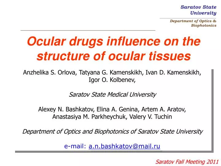
Ocular drugs influence on the structure of ocular tissues. Anzhelika S. Orlova, Tatyana G. Kamenskikh, Ivan D. Kamenskikh, Igor O. Kolbenev, Saratov State Medical University Alexey N. Bashkatov, Elina A. Genina, Artem A. Aratov, Anastasiya M. Parkheychuk, Valery V. Tuchin

E N D
Ocular drugs influence on the structure of ocular tissues Anzhelika S. Orlova, Tatyana G. Kamenskikh, Ivan D. Kamenskikh, Igor O. Kolbenev, Saratov State Medical University Alexey N. Bashkatov, Elina A. Genina, Artem A. Aratov, Anastasiya M. Parkheychuk, Valery V. Tuchin Department of Optics and Biophotonics of Saratov State University e-mail: a.n.bashkatov@mail.ru
Motivation: It is well known that diagnostics and treatment of many diseases of human eye is connected with monitoring of glucose content. However, in spite of numerous investigations dealing with the transport of the metabolite within biological tissue, the problem of monitoring of structural changes of eye cornea under action of glucose has not been studied in detail. Goal of the study is investigation of influence of 40% aqueous glucose solution on stuctural changes in human eye cornea at the noninflamation cornea edema afterpost-surgical phacoemulsification of aging cataract
Materials and Methods: in vitro study The study has been performed with ten samples of rabbit cornea immersed in solutions with different pH in the range from 3 to 9. The buffer solutions have been prepared using sodium citrate (C6H5O7Na25*5H2O), citric acid (C6H8O7*H2O), (Na2HPO4), (KH2PO4), sodium carbonate (Na2CO3), and sodium bicarbonate (NaHCO3). pH of these solutions (3.07, 4.04, 4.91, 5.5, 5.91, 7.01, 8.59, 8.99) have been measured by pH-meter "HANNA" (Portugal) using standard procedure.
Materials and Methods: in vitro study For investigation of hydration degree, the samples have been weighed using electronic balance (SCIENTECH, SA210) with the precision of 1 mg. The tissue samples have been immersed into cuvettes with the buffer solutions. Every 10 min during 2 hours, each sample has been taken out from the solution and weighted. The hydration degree of the cornea samples has been calculated using the equation: where M is the mass of tissue sample and t is time.
Materials and Methods: in vivo study Cornea structure has been investigated using Heidelberg Retina Tomograph (HRT) with the Rostok Cornea Module (RCM) (Heidelberg Engineering GmbH, Germany)
Materials and Methods: in vivo study Cornea thickness has been measured with Topcon 3D OCT-1000 Optical coherence Tomograph (OCT) (Japan)
Materials and Methods: in vivo study Sixty volunteers were enrolledfor the study. The subjects were divided into two groups. The I group included thirty volunteers(21 male and 9 female at the age from 56 to 72 years) with post-surgical edema of cornea after phacoemulsification of aging cataract. The II (control) group included thirty health volunteers (16 male and 14 female at the age from 22 to 30 years). For all persons the study of corneal structure and thickness was performed before the instillation of 40%-glucose solution(2 drops 0.05 ml = 0.1 ml) and in 20 and 40 minutes after the procedure with HRT and OCT
Results: in vitro study The comparison of hydration degree of rabbit cornea and collagen (gelatin) under action of solutions with different pH measured in different time. M is the mass of the sample; t is the time of action. The data about gelatin swelling have been obtained from Lykov A.V. Transport phenomena in capillaro-porous media / Moscow, 1954
Results: in vivo study a b c The typical OCT images obtained from cornea in normal state under action of 40%-glucose solution. (a) – before glucose instillation; (b) – after 20 min; (c) – after 40 min
Results: in vivo study The hydration degree of human cornea with edema measured in vivo under action of 40%-glucose solution (pH = 3.5) in different time The thickness of human cornea measured in vivo by OCT under action of 40%-glucose solution (pH = 3.5) in different time
Results: in vivo study The typical HRT images obtained from corneum epithelium in normal state under action of 40%-glucose solution. (a) – before glucose instillation; (b) – after 20 min; (c) – after 40 min a b c The typical HRT images obtained from corneum epithelium with edema under action of 40%-glucose solution. (a) – before glucose instillation; (b) – after 20 min; (c) – after 40 min
Results: in vivo study The typical HRT images obtained from corneum endothelium in normal state under action of 40%-glucose solution. (a) – before glucose instillation; (b) – after 40 min a b The typical HRT images obtained from corneum endothelium with edema under action of 40%-glucose solution. (a) – before glucose instillation; (b) – after 40 min
Results: in vivo study The diameter of the corneal endothelium cells measured in vivo by HRT under action of 40%-glucose solution in different time The diameter of the corneal epithelial cells measured in vivo by HRT under action of 40%-glucose solution in different time
Acknowledgement: Grant #224014 Network of Excellence for Biophotonics (PHOTONICS4LIFE) of the Seventh Framework Programme of Commission of the European Communities Russian Federation governmental contacts 02.740.11.0770, and 02.740.11.0879 The authors thank Dr. Alexander B. Pravdin (Department of Optics and Biophotonics of Saratov State University) for the help in preparation of the buffer solutions