Download
1 / 27
290 likes | 497 Views
Regulation of Glomerular Filtration. Three mechanisms control the GFR Renal autoregulation (intrinsic system) Neural controls Hormonal mechanism (the renin-angiotensin system). Intrinsic Controls.

E N D
Regulation of Glomerular Filtration • Three mechanisms control the GFR • Renal autoregulation (intrinsic system) • Neural controls • Hormonal mechanism (the renin-angiotensin system)
Intrinsic Controls • Under normal conditions, renal autoregulation maintains a nearly constant glomerular filtration rate
Extrinsic Controls • When the sympathetic nervous system is at rest: • Renal blood vessels are maximally dilated • Autoregulation mechanisms prevail
Extrinsic Controls • Under stress: • Norepinephrine is released by the sympathetic nervous system • Epinephrine is released by the adrenal medulla • Afferent arterioles constrict and filtration is inhibited • The sympathetic nervous system also stimulates the renin-angiotensin mechanism
Renin-Angiotensin Mechanism • Is triggered when the JG cells release renin • Renin acts on angiotensinogen to release angiotensin I • Angiotensin I is converted to angiotensin II • Angiotensin II: • Causes mean arterial pressure to rise • Stimulates the adrenal cortex to release aldosterone • As a result, both systemic and glomerular hydrostatic pressure rise
Renin Release • Renin release is triggered by: • Reduced stretch of the granular JG cells • Stimulation of the JG cells by activated macula densa cells • Direct stimulation of the JG cells via 1-adrenergic receptors by renal nerves • Angiotensin II
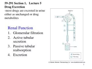
Tubular Reabsorption • All organic nutrients are reabsorbed • Water and ion reabsorption is hormonally controlled • Reabsorption may be an active (requiring ATP) or passive process
Nonreabsorbed Substances • A transport maximum (Tm): • Reflects the number of carriers in the renal tubules available • Exists for nearly every substance that is actively reabsorbed • When the carriers are saturated, excess of that substance is excreted
Nonreabsorbed Substances • Substances are not reabsorbed if they: • Lack carriers • Are not lipid soluble • Are too large to pass through membrane pores • Urea, creatinine, and uric acid are the most important nonreabsorbed substances
Atrial Natriuretic Peptide Activity • ANP reduces blood Na+ which: • Decreases blood volume • Lowers blood pressure • ANP lowers blood Na+ by: • Acting directly on medullary ducts to inhibit Na+ reabsorption • Counteracting the effects of angiotensin II • Indirectly stimulating an increase in GFR reducing water reabsorption
Tubular Secretion • Essentially reabsorption in reverse, where substances move from peritubular capillaries or tubule cells into filtrate • Tubular secretion is important for: • Disposing of substances not already in the filtrate • Eliminating undesirable substances such as urea and uric acid • Ridding the body of excess potassium ions • Controlling blood pH
Formation of Dilute Urine • Filtrate is diluted in the ascending loop of Henle • Dilute urine is created by allowing this filtrate to continue into the renal pelvis • This will happen as long as antidiuretic hormone (ADH) is not being secreted
Formation of Dilute Urine • Collecting ducts remain impermeable to water; no further water reabsorption occurs • Sodium and selected ions can be removed by active and passive mechanisms • Urine osmolality can be as low as 50 mOsm (one-sixth that of plasma)
Formation of Concentrated Urine • Antidiuretic hormone (ADH) inhibits diuresis • This equalizes the osmolality of the filtrate and the interstitial fluid • In the presence of ADH, 99% of the water in filtrate is reabsorbed
Formation of Concentrated Urine • ADH-dependent water reabsorption is called facultative water reabsorption • ADH is the signal to produce concentrated urine • The kidneys’ ability to respond depends upon the high medullary osmotic gradient
Diuretics • Chemicals that enhance the urinary output include: • Any substance not reabsorbed • Substances that exceed the ability of the renal tubules to reabsorb it • Substances that inhibit Na+ reabsorption
Diuretics • Osmotic diuretics include: • High glucose levels • carries water out with the glucose • Alcohol • inhibits the release of ADH • Caffeine and most diuretic drugs • inhibit sodium ion reabsorption • Lasix and Diuril • inhibit Na+-associated symporters
Ureters • Slender tubes that convey urine from the kidneys to the bladder • Ureters enter the base of the bladder through the posterior wall • This closes their distal ends as bladder pressure increases and prevents backflow of urine into the ureters
Ureters • Ureters have a trilayered wall • Transitional epithelial mucosa • Smooth muscle muscularis • Fibrous connective tissue adventitia • Ureters actively propel urine to the bladder via response to smooth muscle stretch
Urinary Bladder • Smooth, collapsible, muscular sac that stores urine • It lies retroperitoneally on the pelvic floor posterior to the pubic symphysis • Males – prostate gland surrounds the neck inferiorly • Females – anterior to the vagina and uterus • Trigone – triangular area outlined by the openings for the ureters and the urethra • Clinically important because infections tend to persist in this region
Urinary Bladder • The bladder wall has three layers • Transitional epithelial mucosa • A thick muscular layer • A fibrous adventitia • The bladder is distensible and collapses when empty • As urine accumulates, the bladder expands without significant rise in internal pressure
Urethra • Muscular tube that: • Drains urine from the bladder • Conveys it out of the body
Urethra • Sphincters keep the urethra closed when urine is not being passed • Internal urethral sphincter • involuntary sphincter at the bladder-urethra junction • External urethral sphincter • voluntary sphincter surrounding the urethra as it passes through the urogenital diaphragm • Levator ani muscle • voluntary urethral sphincter
Female Urethra • The female urethra is tightly bound to the anterior vaginal wall • Its external opening lies anterior to the vaginal opening and posterior to the clitoris
Male urethra • The male urethra has three named regions • Prostatic urethra • runs within the prostate gland • Membranous urethra • runs through the urogenital diaphragm • Spongy (penile) urethra • passes through the penis and opens via the external urethral orifice
Micturition (Voiding or Urination) • The act of emptying the bladder • Distension of bladder walls initiates spinal reflexes that: • Stimulate contraction of the external urethral sphincter • Inhibit the detrusor muscle and internal sphincter (temporarily)
Micturition (Voiding or Urination) • Voiding reflexes: • Stimulate the detrusor muscle to contract • Inhibit the internal and external sphincters