Download
1 / 45
550 likes | 990 Views
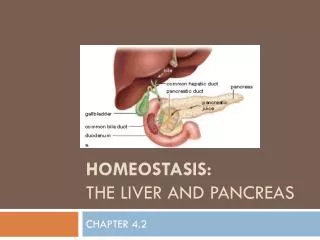
Homeostasis: the liver and pancreas. CHAPTER 4.2. Overview. Mammalian Liver Anatomy Functions Lipid Regulation Protein and Amino Acid Regulation Blood Sugar regulation Bile production Other functions Chemical classification of Hormones Water and lipid soluble hormones

E N D
Homeostasis:the liver and pancreas CHAPTER 4.2
Overview • Mammalian Liver • Anatomy • Functions • Lipid Regulation • Protein and Amino Acid Regulation • Blood Sugar regulation • Bile production • Other functions • Chemical classification of Hormones • Water and lipid soluble hormones • Hormonal Feedback loop • Antagonistic Hormones • Mammalian Pancreas • Blood sugar regulation
Liver: Bodily Metabolic Centre • Largest gland in the body with many metabolic and regulatory roles • Lies on the upper right section of the abdominal cavity, under the diaphragm • Receives plentiful blood supply where substances are extracted for processing. 2 main vessel:- • Hepatic artery – brings oxygenated blood from dorsal artery • Hepatic portal vein – bring nutrient rich blood from small intestines • Hepatic vein - Blood from liver is brought back to heart via this vein and posterior vena cava. • Liver cell/hepatocytes are undifferentiated and structurally identical.
Contains approximately 0.1 millions lobules that serve as structural and functional units • Each lobe contains rows of liver cells 1. Liver artery arm 2. Bile duct 3. Bile duct arm 4. Portal vein a. Lobe (simplified) b. Hepatocyte
Functions of the Liver • Over 500 functions. Most importantly • Regulation of lipids • Lipids used for energy for cellular functions (more energy than glucose) • Liver responsible for proper lipid concentrations in the blood. • Lipids removed from blood by liver cells or transported to fat storage areas in the form of adipose tissue or lipoproteins for brain and nerve tissue synthesis • Cholesterols removed and some converted to bile salts • Fatty acids conversion to acetyl-coA via fatty acid spiral/lipolysis • Lipid synthesis – cholesterol synthesis (Mevalonate pathway) and lipogenesis • Abnormally high lipids – arthrosclerosis, coronary thrombosis
Functions of Liver • Regulation of amino acids and proteins • Non-essential aa can be synthesised by transamination • Excess aa and proteins cannot be stored in the body. Any excess in returned to the liver for catabolism through deamination into non-nitrogenous and nitrogenous parts (amino group - NH2) • The non-nitrogenous, keto acid is converted into glucose in the liver to be stored as glycogen or broken down to release heat. • The nitrogenous ammonia, is potently toxic. Hence, it is converted into urea using the urea or ornithine cycle • This is transported by the blood the kidneys for excretion
Functions of Liver • Bile production • Bile comprises of bile salts and bile pigments that is stored in the gall bladder till needed in fat digestion • Bile salts are synthesized from cholesterol • Bile pigments (yellowish-green) are from the incorporation of the by-products of red blood cell disassembly • Gallstones are an accumulation of cholesterol crystals that can cause blockage of the bile/biliary duct and increase pressure of the gall bladder. • Accumulation stems from bile constituents’ imbalance.
Functions of Liver • Regulation of blood sugar level • Excess glucose is either converted by pancreatic insulin for storage as glycogen; or broken down into H2O + CO2 + heat • When the body has excess glucose, glycogenesis is the synthesis of glycogen from glucose that is stimulated in the presence of the pancreatic hormone insulin. • Prevention of glucose from falling below the crucial level is performed by a process called glycogenolysis. • Glycogenolysis is the catabolism of glycogen that requires the activation of hepatic enzyme glycogen phosphorylase by pancreatic hormone glucagon.
Hexokinase is stimulated by insulin • Glycogen phosphorylase is stimulated by glucagon
Functions of Liver • Regulation of blood sugar level • In skeletal muscles, glycogen cannot be converted into glucose directly through glucose-6-phosphate route as in the liver due to the lack of the enzyme glucose-6-phosphotase. • Instead it is channeled through glycolysis and converted into pyruvate. Consecutively, processed through aerobiosis or anaerobiosis. • In anaerobiosis, lactate that will be carried to the liver for conversion – firstly, to glucose and then glycogen using the Cori cycle.
Functions of Liver • Thermoregulation • Liver is large • Plenty of blood and high metabolic rate • Hence, easy to release excess heat to maintain body temperature. • Detoxification of blood – Kupffer cells • Elimination of steroids • Storage of blood • Formation of red blood cells in foetus • Production of plasma protein (fibrinogen, albumin and globulin) • Storage of vitamins and minerals
Chemical Classes of Hormones • Three major classes of molecules function as hormones in vertebrates: • Polypeptides (proteins and peptides) • Amines derived from amino acids • Steroid hormones • Lipid-soluble hormones (steroid hormones) pass easily through cell membranes, while water-soluble hormones (polypeptides and amines) do not • The solubility of a hormone correlates with the location of receptors inside or on the surface of target cells
Fig. 45-3 Water-soluble Lipid-soluble 0.8 nm Polypeptide: Insulin Steroid: Cortisol Amine: Epinephrine Amine: Thyroxine
Cellular Response Pathways • Water and lipid soluble hormones differ in their paths through a body • Water-soluble hormones are secreted by exocytosis, travel freely in the bloodstream, and bind to cell-surface receptors • Lipid-soluble hormones diffuse across cell membranes, travel in the bloodstream bound to transport proteins, and diffuse through the membrane of target cells
Fig. 45-5-2 Fat-soluble hormone Water- soluble hormone Transport protein Signal receptor TARGET CELL OR Signal receptor Cytoplasmic response Gene regulation Cytoplasmic response Gene regulation NUCLEUS (a) (b)
Negative feedback and antagonistic hormone pairs • Hormones are assembled into regulatory pathways • Hormones are released from an endocrine cell, travel through the bloodstream, and interact with the receptor or a target cell to cause a physiological response • A negative feedback loop inhibits a response by reducing the initial stimulus • Negative feedback regulates many hormonal pathways involved in homeostasis
Fig. 45-11 Pathway Example – Stimulus Low pH in duodenum S cells of duodenum secrete secretin ( ) Endocrine cell Negative feedback Blood vessel Target cells Pancreas Bicarbonate release Response
Insulin and Glucagon: Control of Blood Glucose • Insulin and glucagon are antagonistic hormones that help maintain glucose homeostasis • Glucose that is absorbed from the gut into the hepatic portal vein, increases the blood glucose concentration. This is detected by the pancreas • The pancreas has clusters of endocrine cells called islets of Langerhans with alpha cells that produce glucagon and beta cells that produce insulin
Pancreas: Endo- and Exocrine Functions • Lies deep within the abdominal cavity, on the posterior of the abdominal wall • Elongated and somewhat flattened organ with endo- and exocrine functions. • As an exocrine gland, it functions in the digestive system due to the secretion of pancreatic juice via the ducts to the small intestines. • As an endocrine gland, it function in the secretion of hormones (insulin, glucagon and somatostatin) • This is thanks to the cells on the islet of Langerhans
Fig. 45-12-1 Insulin Beta cells of pancreas release insulin into the blood. STIMULUS: Blood glucose level rises. Homeostasis: Blood glucose level (about 90 mg/100 mL)
Fig. 45-12-2 Body cells take up more glucose. Insulin Beta cells of pancreas release insulin into the blood. Liver takes up glucose and stores it as glycogen. STIMULUS: Blood glucose level rises. Blood glucose level declines. Homeostasis: Blood glucose level (about 90 mg/100 mL)
Fig. 45-12-3 Homeostasis: Blood glucose level (about 90 mg/100 mL) STIMULUS: Blood glucose level falls. Alpha cells of pancreas release glucagon. Glucagon
Fig. 45-12-4 Homeostasis: Blood glucose level (about 90 mg/100 mL) STIMULUS: Blood glucose level falls. Blood glucose level rises. Alpha cells of pancreas release glucagon. Liver breaks down glycogen and releases glucose. Glucagon
Fig. 45-12-5 Body cells take up more glucose. Insulin Beta cells of pancreas release insulin into the blood. Liver takes up glucose and stores it as glycogen. STIMULUS: Blood glucose level rises. Blood glucose level declines. Homeostasis: Blood glucose level (about 90 mg/100 mL) STIMULUS: Blood glucose level falls. Blood glucose level rises. Alpha cells of pancreas release glucagon. Liverbreaks downglycogen andreleases glucose. Glucagon
Target Tissues for Insulin and Glucagon • Insulin reduces blood glucose levels by • Promoting the cellular uptake of glucose • Slowing glycogen breakdown in the liver • Promoting fat storage (lipogenesis) • Glucagon increases blood glucose levels by • Stimulating conversion of glycogen to glucose in the liver • Stimulating breakdown of fat and protein into glucose
Diabetes Mellitus • Diabetes mellitus is perhaps the best-known endocrine disorder • It is the failure of glucose homeostasis • It is caused by a deficiency of insulin or a decreased response to insulin in target tissues • It is marked by elevated blood glucose levels
Diabetes Mellitus • Type I diabetes mellitus (insulin-dependent) (30%) is an autoimmune disorder in which the immune system destroys pancreatic beta cells • Type II diabetes mellitus (non-insulin-dependent) (70%) involves insulin deficiency or reduced response of target cells due to change in insulin receptors
You should now be able to: • Note the anatomy and function of the liver lobules and their components • Difference in canaliculi and sinusoid. • Distinguish between the major functions of the liver especially lipid, protein, amino acids and glucose regulation. • Describe the difference between water-soluble and lipid-soluble hormones • Explain how the antagonistic hormones insulin and glucagon regulate carbohydrate metabolism • Distinguish between type 1 and type 2 diabetes