Hyperplasia
Hyperplasia. Dr : Hala El-sayed Mahmoud Lecturer of pathology. Hyperplasia. Definition; Hyperplasia is an increase in the number of cells in an organ or tissue, usually resulting in increased volume of the organ or tissue. Physiologic Hyperplasia : can be divided into:

Hyperplasia
E N D
Presentation Transcript
Hyperplasia Dr : Hala El-sayed Mahmoud Lecturer of pathology
Hyperplasia Definition; Hyperplasia is an increase in the number of cells in an organ or tissue, usually resulting in increased volume of the organ or tissue. Physiologic Hyperplasia: can be divided into: (1) Hormonal hyperplasia, Examples, hyperplasia of the female breast at puberty (2)Compensatory hyperplasia, Examples, after partial hepatectomy
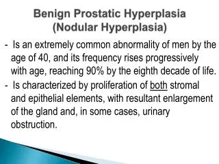
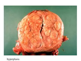
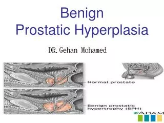
Pathologic Hyperplasia Mostly caused by excessive hormonal stimulation or growth factors acting on target cells. Examples: Endometrial hyperplasia is an example of abnormal hormone-induced hyperplasia, due to increases in estrogen, Benign prostatic hyperplasia, due to abnormal response to androgens. Hyperplasia of lymphoid tissue in response to antigenic stimulation. NB; although these forms of hyperplasia are abnormal, the process remains controlled, because the hyperplasia regresses if the hormonal stimulation is eliminated. Hyperplasia
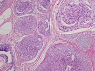
Section in the prostate shows: Increase in the number of acini. They are variable in size and shape, lined by columnar cells with intraluminal papillary formations. Some acini are cystically dilated and lined by flattened cells. The lumen of the acini contain shedded epithelial cells or rounded homogenous pink corpora amylacae. The fibromuscular stroma is increased with lymphocytic infiltrate. Diagnosis: Senile prostatic hyperplasia
Corpora amylacea Prostatic hyperplasia