Integumentary System
410 likes | 436 Views
Learn about the different components of the integumentary system, such as the skin, hair, glands, nails, and nerve endings. Discover the anatomy, physiology, and functions of the system. Explore the layers of the epidermis and the importance of the dermis. Gain knowledge about thin and thick skin and their differences.

Integumentary System
E N D
Presentation Transcript
This system is divided into: 1- skin 2- hair 3- glands 4- nails 5- nerve endings I) Skin Skin is an organ because it consists of different tissues that are joined to perform a specific function. Largest organ of the body in surface area and weight. Dermatology is the medical specialty concerning the diagnosing and treatment of skin disorders.
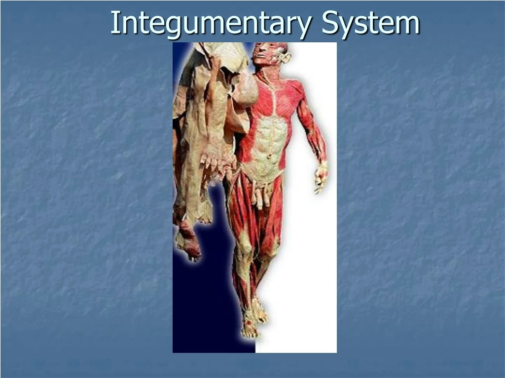
Anatomy (structure) Epidermis (thinner outer layer of skin) Dermis (thicker connective tissue layer) Hypodermis (subcutaneous layer or Sub-Q) Muscle and bone Physiology (function) 1- Protection - physical barrier that protects underlying tissues from injury, UV light and bacterial invasion. - mechanical barrier is part non specific immunity (skin, tears and saliva).
2- Regulation of body temperature - high temperature or strenuous exercise; sweat is evaporated from the skin surface to cool it down. - vasodilation (increases blood flow) and vasoconstriction (decrease in blood flow) regulates body temp. 3-Sensation - nerve endings and receptor cells that detect stimuli to temp., pain, pressure and touch.
4- Excretion - sweat removes water and small amounts of salt, uric acid and ammonia from the body surface 5- Blood reservoir - dermis houses an extensive network of blood vessels carrying 8-10% of total blood flow in a resting adult. 6- Synthesis of Vitamin D (cholecalciferol) -UV rays in sunlight stimulate the production of Vit. D. Enzymes in the kidney and liver modify and convert to final form; calcitriol (most active form of Vit. D.) Calcitriol aids in absorption of calcium from foods and is considered a hormone.
Epidermis: keratinized stratified squamous epithelium with four distinct cell types and five distinct layers.
Cells in the epidermis: - keratinoytes - melanocytes - Merkel cells - Langerhans’ cells 1- Keratinocytes: most abundant - produce keratin (fibrous protein) - protective; waterproofing the skin - continuous mitosis - form in the deepest layer called the stratum basale - cells push their way up to the surface where they are dead cells filled with keratin; will slough off. Regenerates every 25-45 days.
2- Melanocytes: - cells produce brownish/black pigment called melanin. (8% of epidermal cells) - stratum basale - branching processes (dendrites) - melanin accumulates in melanosomes and transported along dendrites of the melanocytes to keratinocytes. - melanin accumulates on the superficial aspect of the keratinocyte shielding its nucleus from harmful UV light. - lack of melanin: albino
3- Merkel cells: - stratum basale - epidermis of hairless skin - attach to keratinocytes by desmosomes - make contact with a sensory neuron ending called a Merkel disc (touch). 4- Langerhans’ cells: - star-shaped cells arising from bone marrow that migrate to epidermis. - epidermal dendritic cells (macrophages) - interact with a WBC called a T- helper cell - easily damaged by UV light.
Stratum corneum Stratum lucidum Stratum granulosum Stratum spinosum Stratum basale
Differences between thin & thick skin Thin Skin Thick Skin Entire body except thick skin areas. Less than 5 layers of stratum corneum with no stratum lucidum Hair follicles present except lips, labia minora, and glans penis • Palms of hands and soles of feet = acral skin • 5 layers thick stratum corneum with increased granular layer • More sensory receptors • Lack sebaceous glands and increased eccrine glands • No hair follicles
5 layers of the epidermis: 1- Stratum corneum (horny layer) - layer has many rows of dead cells filled with keratin - continuously shed and replaced (desquamation) - effective barrier against light, heat and bacteria - 20-30 cell layers thick - dandruff and flakes - 40 lbs. of skin flakes in a lifetime (dust mites!)
2- Stratum lucidum -seen in thick skin of the palms and soles of feet. - 3-5 rows of clear flat dead cells - keratohyalin (precursor) to keratin 3- Stratum granulosum - 3-5 rows of flattened cells - nuclei of cells flatten out - organelles disintegrate cells eventually die - keratohyalin granules (darkly stained) accumulate - lamellated granules secrete glycolipids into extracellular spaces to slow water loss in the epidermis
4- Stratum spinosum: “spiny layer” - 8-10 rows of polyhedral (many sided) cells - appearance of prickly spines - shrink when prepared for slide - melanin granules and Langerhans’ cell predominate
5- Stratum basale: deepest epidermal layer - attached to dermis - single row of cells - mostly columnar keratinocytes - with rapid mitotic division - stratum germinativum - contain merkel cells and melanocytes - 10-25%
Dermis • Two layers • Papillary dermis = includes the dermal papilla which project into the epidermis • The increases contact area preventing epidermal detachment • Also results in an undulating pattern which vary by anatomic location and individual resulting in grooves in the epidermis =dermatoglyphics (fingerprints) • Capillaries, free nerve endings and encapsulated sensory receptors called Meissner’s corpuscles. • Reticular dermis = area between the papillary dermis and subcutis
Dermis: - flexible and strong connective tissue - elastic, reticular and collagen fibers - cells: fibroblasts, macrophages (WBC), mast cells (histamine). - nerves, blood and lymphatic vessels - oil and sweat glands originate - two layers: papillary and reticular
1- Papillary layer: - loose connective tissue with nipple like surface projection called dermal papilla. - capillaries - contain pain receptors - contain touch receptors (Meissner’s corpuscles - dermal ridges- epidermal ridges- pattern called fingerprints
2- Reticular layer: - dense irregular c.t. - collagen fibers offer strength - holds water - dermal tearing causes stretch marks. - striae Skin color: attributed to melanin, hemoglobin and carotene. Race is determined by amount of melanin not # of melanocytes.
Hemoglobin (blood) will impart pinkish tones to skin. Blushing 1- Redness (erythema) - reddened skin, embarrassment, fever, hypertension, inflammation, or allergy 2- Pallor/blanching - pale skin, emotional distress or anemia, low blood pressure 3- Jaundice - liver disease, bile deposited in tissue 4- Bronzing - bronze coloration (Addison's disease) hypofunction of adrenal cortex 5- Black & blue - bruises, escaped blood clots in tissue spaces (clotted blood masses = hematomas)
Hair color: Dark hair: mostly melanin Blond and red hair: melanin with Fe and S. Gray hair: loss of pigment (decr. tyrosinase) White hair: air bubbles in the medullary hair shaft.
Hair (pili) - main function is protection - hair root nerve plexus for touch - normal hair loss in adult 70-100 hairs/day
Hair anatomy: - composed of dead columns of keratinized cells. - shaft: is the superficial portion of hair - root: below the surface in the dermis Shaft and root are composed of three layers: inner medulla, middle cortex and outer cuticle. Inner medulla has 2-3 rows of polyhedral cells where pigment is located Cortex is major portion of shaft Cuticle is scaly and heavily keratinized (shingles)
Vellus hair: fine hair Terminal hair : coarser hair; axillary and pubic region. Grow in response to sex hormonesHirsutism: excessive hairiness: incr. androgens
Hair follicle surrounds the root. Bulb is the enlargement at the end of the follicle. - Also houses the germinal layer Papilla (nipple like) is located in the bulb and is where the blood supply nourishes the hair.
Glands: Two types of glands exist in the integument. - Sebaceous glands (oil glands) - Sudoriferous glands (sweat glands) Sebaceous glands: (holocrine glands) - connected to hair follicle - not found on palms and soles of feet - secretes sebum (fats, cholesterol and proteins - keep hair from drying out, keeps skin moist - whiteheads, blackheads and acne
Nails: - Produced by cells in the epidermis - Nail plate (body): visible portion - Nail root: located under cuticle - Lunula: half moon crescent shaped white portion under cuticle - Nail bed: located under nail plate - Hypoxia: decr. oxygen in blood, nail bed will turn blue- cyanosis


