Download

1 / 8
100 likes | 304 Views
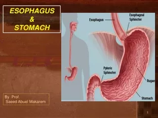
Imaging of esophagus and stomach. Radiology department Dr. A. Alhawas. 3. 1. 2. 4. Splenic artery Abdominal aorta Common hepatic artery Gastro-duodenal artery. Abdominal aorta. Common hepatic artery. Splenic artery. Gasto -duodenal artery. Left gasto-epiploic artery.

E N D
Imaging of esophagus and stomach Radiology department Dr. A. Alhawas
3 1 2 4 Splenic artery Abdominal aorta Common hepatic artery Gastro-duodenal artery
Abdominal aorta. • Common hepatic artery. • Splenic artery. • Gasto-duodenal artery. • Left gasto-epiploic artery.
Left main pulmonary artery. • Esophagus. • Thoracic arota. • Right main pulmonary artery. • Left main bronchus. • Azygousvien. 1 4 5 2 6 3
Stomach • Splenic artery / short gastric artery. • Gastro-esophageal junction. • Inferior vena cava. • Aorta. 1 3 4 2 5
State which layers are pathologically changed in pyloric stenosis. • both circular and longitudinal layers . • State the name of arteries supplying the pylorus and the first part of the duodenum. • State four (4) structures forming the stomach bed: • Pancreas ( body and tail ). • Spleen. • Left kidney. • Left adrenal gland. • Left crus of the diaphragm. • Splenic artery • Transverse mesocolon.