Download
1 / 44
910 likes | 2.07k Views
Principles of Neuronavigation: Frame and Frameless. History. “Stereotactic”: From Greek “stereos”=3-dimensional and Latin “tactus”=to touch

E N D
History • “Stereotactic”: From Greek “stereos”=3-dimensional and Latin “tactus”=to touch • 1908: Horsely and Clarke develop first apparatus for insertion of probes into the brain based upon Cartesian planes and bony landmarks; only used in primates • 1947: Spiegel and Wycis report first human use of stereotactic device . Goal was to perform minimally-invasive psychosurgery but first use was movement disorders • 1948: Leksell develops first arc-centered frame • 1957: Talairach publishes first atlas based upon ventriculography and intracranial brain landmarks rather than bone landmarks • 1986: Kelley develops frame-based system for eye-tracking of operative microscope • 1986: Roberts develops frameless acoustic-based system for tracking operative microscope • 1991: Bucholz develops the first prototype for frameless sonic navigation of tracking tools and instruments in human cranial surgery • Soon after incorporated optical digitizers to reduce inaccuracies from sound echoes
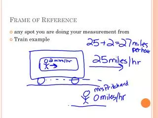
General Principles of Stereotaxy • Navigation is based upon targeting relative to known reference points • Fiducial : • From latin “fiducia” meaning trust • A point of reference that can be visualized on imaging and identified by the surgeon and/or software package • Accuracy of targeting is influenced by the number of fiducials around a target zone and the constancy of fiducials relative to the target • Frame-based stereotaxy: Fiducials are bars built into cage or box that sits on frame during imaging • Frameless stereotaxy: Fiducials are reference markers (stickers, bone screws) which are fixed directly to the patient prior to imaging
Co-registration is the fundamental principle of stereotaxy 1906 -- Horsley & Clarke (animal) stereotactic frame
Co-registration is the fundamental principle of stereotaxy 1906 – Horsley & Clarke (animal) stereotactic frame 1947 – Spiegel & Wycis (human) stereotactic frame
Co-registration is the fundamental principle of stereotaxy 1906 – Horsley & Clarke (animal) stereotactic frame 1947 – Spiegel & Wycis (human) stereotactic frame 1947-1980 – Proliferation of stereotactic frames
Co-registration is the fundamental principle of stereotaxy 1906 – Horsley & Clarke (animal) stereotactic frame 1947 – Spiegel & Wycis (human) stereotactic frame 1947-1980 – Proliferation of stereotactic frames 1980s – Computational resources enable “frameless” transformation-based stereotactic systems
Considerations with Frame-Based Stereotaxy • Method of target localization • Indirect vs. direct • Imaging errors due to frame placement • Imaging errors due to distortion
Methods of Image-Based Target Localization • Indirect (Based upon position of AC-PC) • Standard coordinates • Leksell’s pallidotomy target is classic example • Adjusted map • Schaltenbrand-Wahren is most common • Average AC-PC distance is 23-27mm; greater than 30mm should raise accuracy concerns • Direct (Target visually chosen from scan)
AC-PC: Sagittal T2 Localizer PC Colliculi AC
Indirect Targeting: Fixed Coordinates • Thalamus (Vim) • 1-7mm posterior • 0-3mm superior • 12-17mm lateral • GPi • 2-3mm anterior • 3-6mm inferior • 18-22 mm lateral • STN • 3-5mm posterior • 5-6mm inferior • 11-14mm lateral (All points relative to midcommissural point)
Sources of Error: MRI Image Distortion • Magnetic field inhomogeneities and non-linear magnetic field gradients cause distortion • Distortion often worst in coronal sections; measuring Leksell fiducials can determine distortion severity • Frame may introduce additional distortion • Measuring target distance from MCP on preop MRI can guide targeting from framed image • CT not subject to these distortions; CT/MRI fusion may minimize effects of distortion • Bandwidth can influence contrast • Lower bandwidth increases gray/white contrast to a point • Very low bandwidth can worsen distortion
Eight Things Every Neurosurgery Resident Should Know about Frameless Image-Guidance
What is image-guided surgery and how does it work? Image-guided surgery (neuronavigation, “frameless stereotaxy”) is an operative technique by which correlation between imaging studies and the operative field is provided. This is accomplished by co-registration of imaging studies with the OR patient.
Image overlay, 2I 2ITM Polaris Microscope calibration Microscope frame, M Preop. 3D image, 3I Head in the world space, W WT3I MTW Patient registration Tracking patient and microscope Matrix Expression P2I = 2ITMMTWWT3I P3I
What equipment is involved? • Localization device (digitizer) • e.g., optical, electromagnetic, articulated arm • most systems today include a reference frame to enable OR table movement
What equipment is involved? • Localization device (digitizer) • e.g., optical, electromagnetic, articulated arm • Computer with registration algorithm
What equipment is involved? • Localization device (digitizer) • e.g., optical, electromagnetic, articulated arm • Computer with registration algorithm • Effector • e.g., pointer and monitor, microscope heads-up display
What types of co-registration strategies can be used? Paired-point rigid transformation Surface (contour) matching
Some important definitions… Fiducial registration error (FRE) the root- mean square distance between corresponding fiducial points after registration Fitzpatrick & West, 2001
Fiducial localization error (FLE) the error in locating the fiducial points Fitzpatrick & West, 2001
Target registration error (TRE) the distance between corresponding points other than the fiducial points after registration This is what really matters! Fitzpatrick & West, 2001
Accuracy in phantom testing Benardete et al, 2001
Clinical application accuracy(comparing seven registration methods) Mascott et al, 2006
What are the sources of error? • Imaging data set • resolution • e.g., slice thickness, pixel/voxel size • spatial infidelity • e.g., magnetic field inhomogenieties in echo planar fMRI • imaging study fusion • e.g., CT–MRI, atlas–MRI
Dependence of stereotactic accuracy on image slice thickness Maciunas et al, 1994
What are the sources of error? Sumanaweera, 1994 • Imaging data set • resolution • e.g., slice thickness, pixel/voxel size • spatial infidelity • e.g., magnetic field inhomogenieties in echo planar fMRI
What are the sources of error? • Imaging data set • resolution • e.g., slice thickness, pixel/voxel size • spatial infidelity • e.g., magnetic field inhomogenieties in echo planar fMRI • imaging study fusion • e.g., CT–MRI, atlas–MRI
What are the sources of error? • Imaging data set • Registration process (image–OR space) • axes orientation (handedness of coordinate system) • algorithm ambiguity • fiducial number, configuration, displacement, OR localization (surgeon & digitizer)
Number of fiducials and accuracy Steinmeier et al, 2000 Fitzpatrick et al, 1998
TRE has an approximate N-1/2 dependence Fitzpatrick et al, 1998
Error increases as the distance of the target from the fiducial centroid West et al, 2001
FRE is not a reliable indicator of registration accuracy (!!) Fitzpatrick et al, 1998 FRE is independent of fiducial configuration FRE is independent of bias errors (e.g., MRI gradient, digitizer camera malalignment, bent handheld probe)
Tips regarding fiducials partially adapted from West et al, 2001 Avoid linear fiducial configurations Arrange fiducials so that the center of their configuration is close to the region of interest during surgery Spread out the fiducials Use as many fiducials as reasonably possible Mark scalp at fiducial site Avoid occipital region or distorted scalp
What are the sources of error? Wang & Song, 2011 • Imaging data set • Registration process (image–OR space) • Digitizer performance
What are the sources of error? Dorward et al, 1998 Hill et al, 1998 Surgical field displacement or deformation Roberts et al, 1998 Ji et al, 2012
How does this relate to intraoperative MRI/CT? Numerous implementations Facilitated co-registration Updated image data-set Cost-benefit analyses pending
In what applications has image-guidance been important? Tumor (biopsy, resection of glial and met tumor) Epilepsy (structural & physiologic data, resection) Functional (DBS) Spine (instrumentation) Radiosurgery (frameless technologies) Cerebrovascular (?) Other: ENT, Plastics, Ortho, General
What’s under development for image-guidance? Nathoo, 2005 Louw, 2004 • Automated registration • Ease of use • Updated imaging/registration • Increasing accuracy • Robotics • Extension of application to other • surgeries, other disciplines