Download
1 / 73
730 likes | 885 Views
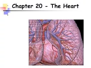
Lecture 20, The Heart . Lecturer: Dr. Barjis Room P313 Phone: (718) 260-5285 E-Mail: ibarjis@citytech.cuny.edu. Learning Objectives. Describe the organization of the cardiovascular system. Describe the location and general features of the heart, including the pericardium.

E N D
Lecture 20, The Heart Lecturer: Dr. Barjis Room P313 Phone: (718) 260-5285 E-Mail: ibarjis@citytech.cuny.edu
Learning Objectives • Describe the organization of the cardiovascular system. • Describe the location and general features of the heart, including the pericardium. • Discuss the differences between nodal cells and conducting cells and describe the components and functions of the conducting system of the heart. • Identify the electrical events associated with a normal electrocardiogram.
Learning Objectives • Explain the events of the cardiac cycle including atrial and ventricular systole and diastole, and relate the heart sounds to specific events in the cycle. • Define cardiac output, heart rate and stroke volume and describe the factors that influence these variables. • Explain how adjustments in stroke volume and cardiac output are coordinated at different levels of activity.
The cardiovascular system is divided into two circuits • Pulmonary circuit • Delivers blood from the right ventricle of the heart to the lungs and from the lungs to the left atrium of the heart • System circuit • Delivers blood from the left ventricle of the heart to the rest of the body and collects blood from the rest of the body and delivers it to the right atrium of the heart.
An Overview of the Cardiovascular System • Pulmonary circuit: • Right ventricle Pulmonary Artery Muscular Arteries Lungs (Arterioles Capillaries Venules) Medium Size Veins Pulmonary veins Left Atrium • System circuit • Left Ventricle Aorta Muscular Arteries Arterioles Capillaries Venules Medium Size Veins Superior and Inferior Vena Cava • Right Atrium
Blood Flow Through the Blood Vessels • Blood flows through the blood vessels from the heart and back to the heart in the following order: • Elastic Arteries e.g. Aorta, pulmonary artery • Muscular Arteries • Arterioles • Capillaries – the only vessels that allow exchange • Venules • Medium Veins • Large Veins e.g. vena cava, pulmonary vein
Anatomy of the Heart The pericardia • Visceral pericardium or epicardium • Parietal pericardium • Pericardial fluid
Superficial Anatomy of the Heart • The heart consists of four chambers • Two upper chamber called atria • Two lower chambers called ventricles • The two upper and two lower chambers are separated by atrioventricular valve
The Heart Wall • The heart wall is composed of three layers: • Epicardium is primarily composed of Areolar Tissue and epithelium • Myocardium is primarily composed of cardiac muscle tissue • Endocardium is primarily composed of Areolar Tissue and endothelium
Internal Anatomy and Organization • Right Atrium • Thin walled chambers that receive blood from superior and inferior vena cava and pumps blood to the right ventricle • Left Atrium • Thin walled chambers that receive blood from pulmonary veins and pumps blood to left ventricle • Right Ventricle • Thick walled chamber that receives blood from right atrium and pumps blood to pulmonary artery. • Left Ventricle • Thick walled chamber that receives blood from left atrium and pumps blood to the Aorta.
Internal Anatomy and Organization • The two ventricles are separated from the atria by atrioventricular (AV) valves • Tricuspid valve separates right atrium from right ventricle • Bicuspid valve separates left atrium from left ventricle • Chordae tendineae • Tendinous fibers attached to the cusps of AV valves • It attaches the cusps of atrioventricular valves to papillary muscles • It prevents the AV valve from reversing into the atria as papillary muscles contract
Blood flow through the heart • Right atria –receives blood from superior and inferior vena cava and pumps it to the right ventricle through the tricuspid valve • Right ventricle –receives blood from right atrium and pumps it toto the pulmonary artery through the pulmonary semilunar valve • Pulmonary artery -delivers the blood to the lungs • At the lungs gas exchange occur • Oxygen diffuses from the alveoli to the capillary and carbon dioxide diffuses from the capillary to the alveoli.
Blood flow through the heart • Pulmonary Vein - after the gas exchange at the lungs pulmonary veins collect the blood and delivers them to the left atrium. • Left atria – receives blood from pulmonary veins and pumps it to the left ventricle through the bicuspid valve • Left ventricle- receives blood from the left atria and pumps it to the aorta through the aortic semilunar valve
Blood flow through the heart • Aorta branches into smaller arteries and delivers the blood to the cells throughout the body. • Gas exchange occur between the cell and the capillaries • Oxygen diffuses from the capillaries to the cell and carbon dioxide diffuses from the cell to the capillaries. • After the gas exchange the blood is delivered back to the heart by superior and inferior vena cava.
Structural Differences in heart chambers and valves • Compared to the right ventricle the left ventricle is: • More muscular and has thicker wall • Develops higher pressure during contraction • Produces about 6 times more force during contraction • Round in cross section • Functions of valves • AV valves prevent backflow of blood from the ventricles to the atria • Semilunar valves prevent backflow of blood from the pulmonary trunk and aorta to the ventricles.
Blood Supply to the Heart • Coronary arteries are the first blood vessels to branch from the aorta • Coronary arteries supply blood to the heart and coronary veins collect the blood from the heart • Arteries include the right and left coronary arteries, marginal arteries, anterior and posterior interventricular arteries, and the circumflex artery • Veins include the great cardiac vein, anterior and posterior cardiac veins, the middle cardiac vein, and the small cardiac vein
The Heartbeat Cardiac Physiology • Two classes of cardiac muscle cells • Specialized muscle cells of the conducting system • Contractile cells
The Conducting System • The conducting system includes: • Sinoatrial (SA) node - Pacemaker cells are located in the SA node • Atrioventricular (AV) node • AV bundle, • bundle branches, and • Purkinje fibers Animation: Heart flythrough (see tutorial)
Impulse Conduction through the heart • SA node begins the action potential • Stimulus spreads to the AV node • Impulse is delayed at AV node • Impulse then travels through ventricular conducting cells • Then distributed by Purkinje fibers
Impulse Conduction through the Heart Animation: Cardiac Activity (see tutorial)
The electrocardiogram (ECG) • ECG is a recording of the electrical events occurring during the cardiac cycle • The P wave of ECG indicates the depolarization of the atriums • The QRS complex of ECG indicates the depolarization of the ventricles • The T wave of ECG indicates ventricular repolarization • Analysis of ECG can reveal • Condition of conducting system • Effect of altered ion concentration • Size of ventricles • Position of the heart
Contractile Cells • Resting membrane potential of approximately –90mV • Action potential • Rapid depolarization • A plateau phase unique to cardiac muscle • Calcium channels remain open longer than the sodium channels • Repolarization • Refractory period follows the action potential
The cardiac cycle • The period between the start of one heartbeat and the beginning of the next • During a cardiac cycle • Each heart chamber goes through systole and diastole • Correct pressure relationships are dependent on careful timing of contractions Animation: Intrinsic Conduction System (see tutorial)
Heart sounds • Auscultation – listening to heart sound via stethoscope • Four heart sounds • S1 – “lubb” caused by the closing of the AV valves • S2 – “dupp” caused by the closing of the semilunar valves • S3 – a faint sound associated with blood flowing into the ventricles • S4 – another faint sound associated with atrial contraction
Cardiodynamics Stroke Volume and Cardiac Output • Stroke volume - is the volume of blood ejected with each ventricle contraction • Cardiac output – is the amount of blood pumped by each ventricle in one minute • Cardiac output equals heart rate times stroke volume
Medulla Oblongata centers affect autonomic innervation • Cardioacceleratory center activates sympathetic neurons • Cardioinhibitory center controls parasympathetic neurons • Medulla Oblongata centers receives input from higher centers, monitoring blood pressure and dissolved gas concentrations
Autonomic Activity • Heart is innervated by sympathetic and parasympathetic nerves. • Sympathetic stimulation • Positive inotropic effect • Releases NE • Parasympathetic stimulation • Negative inotropic effect • Releases ACh
Summary: Regulation of Heart Rate and Stroke Volume • Sympathetic stimulation increases heart rate • Parasympathetic stimulation decreases heart rate • Circulating hormones, specifically E, NE, and T3, accelerate heart rate • Increased venous return increases heart rate • EDV is determined by available filling time and rate of venous return • ESV is determined by preload, degree of contractility, and afterload
Functions and Composition ofBlood • Fluid connective tissue • Functions include • Transporting dissolved gases, nutrients, hormones, and metabolic wastes • Regulating pH and ion composition of interstitial fluids • Restricting fluid loss at injury sites • Defending the body against toxins and pathogens • Regulating body temperature by absorbing and redistributing heat
The Composition of Whole Blood The chief difference between plasma and interstitial fluid involves the concentration of dissolved oxygen and proteins.
Hemopoiesis • Process of blood cell formation • Hemocytoblasts are circulating stem cells that divide to form all types of blood cells • Whole blood from anywhere in the body has roughly the same temperature (38ºC), pH (7.4) and viscosity. • Bright red color if taken from artery • Dull red color if taken from vein
Plasma • Accounts for 46-63% of blood volume • 92% of plasma is water • Higher concentration of dissolved oxygen and dissolved proteins than interstitial fluid
Plasma proteins • more than 90% are synthesized in the liver • Albumins are the most abundant plasma proteins • 60% of plasma proteins • Responsible for viscosity and osmotic pressure of blood
Additional Plasma Proteins • Globulins • ~35% of plasma proteins • Include immunoglobins which attack foreign proteins and pathogens • Include transport globulins which bind ions, hormones and other compounds • Fibrinogen • Converted to fibrin during clotting • Are necessary for blood clotting • Removal of fibrinogen leaves serum
Red Blood Cells Abundance of RBCs • Erythrocytes (RBC) account for slightly less than half the blood volume, and 99.9% of the formed elements. • Hematocrit measures the percentage of whole blood occupied by formed elements • Commonly referred to as the volume of packed red cells
Hemoglobin • Molecules of hemoglobin account for 95% of the proteins in RBCs • Hemoglobin is a globular protein, formed from two pairs of polypeptide subunits • Each subunit contains a molecule of heme which reversibly binds an oxygen molecule • Damaged or dead RBCs are recycled by phagocytes
RBC life span and circulation • Replaced at a rate of approximately 3 million new blood cells entering the circulation per second. • Replaced before they hemolyze • Components of hemoglobin individually recycled • Heme stripped of iron and converted to biliverdin, then bilirubin • Iron is recycled by being stored in phagocytes, or transported throughout the blood stream bound to transferrin
RBC Production • Erythropoeisis = the formation of new red blood cells • Occurs in red bone marrow • Process speeds up with in the presence of EPO (Erythropoeisis stimulating hormone) • RBCs pass through reticulocyte and erythroblast stages